

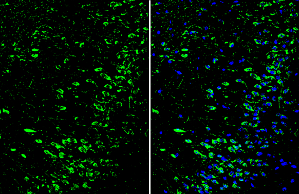
Dopamine Receptor D1 antibody [C2C3], C-term detects Dopamine Receptor D1 protein by immunohistochemical analysis. Sample: Frozen-sectioned mouse cerebral cortex. Green: Dopamine Receptor D1 stained by Dopamine Receptor D1 antibody [C2C3], C-term (GTX100354) diluted at 1:250. Blue: Fluoroshield with DAPI (GTX30920).
 antibody at 1:500 dilution.
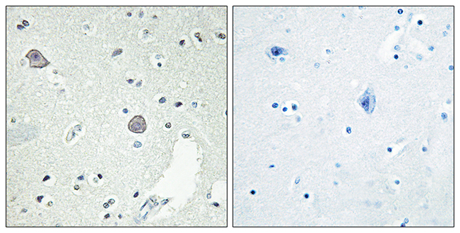
Antigen Retrieval: Trilogy? (EDTA based, pH 8.0) buffer, 15min")
![Dopamine Receptor D1 antibody [C2C3], C-term detects Dopamine Receptor D1 protein by immunofluorescent analysis. Sample: DIV10 rat E18 primary cortical neuron cells were fixed in 4% paraformaldehyde at RT for 15 min. Green: Dopamine Receptor D1 stained by Dopamine Receptor D1 antibody [C2C3], C-term (GTX100354) diluted at 1:500. Red: beta Tubulin 3/ Tuj1, stained by beta Tubulin 3/ Tuj1 antibody [GT11710] (GTX631836) diluted at 1:500. Blue: Fluoroshield with DAPI (GTX30920).](https://www.genetex.com/upload/website/prouct_img/normal/GTX100354/GTX100354_39609_20181227_ICC_IF_R_w_23060100_553.webp "Dopamine Receptor D1 antibody [C2C3], C-term detects Dopamine Receptor D1 protein by immunofluorescent analysis. Sample: DIV10 rat E18 primary cortical neuron cells were fixed in 4% paraformaldehyde at RT for 15 min. Green: Dopamine Receptor D1 stained by Dopamine Receptor D1 antibody [C2C3], C-term (GTX100354) diluted at 1:500. Red: beta Tubulin 3/ Tuj1, stained by beta Tubulin 3/ Tuj1 antibody [GT11710] (GTX631836) diluted at 1:500. Blue: Fluoroshield with DAPI (GTX30920).")
![Dopamine Receptor D1 antibody [C2C3], C-term detects Dopamine Receptor D1 protein by immunofluorescent analysis. Sample: Mock and transfected 293T cells were fixed in ice-cold MeOH for 5 min. Green: Dopamine Receptor D1 stained by Dopamine Receptor D1 antibody [C2C3], C-term (GTX100354) diluted at 1:500. Blue: Fluoroshield with DAPI (GTX30920).](https://www.genetex.com/upload/website/prouct_img/normal/GTX100354/GTX100354_39609_20240927_ICC_IF_B_24100318_597.webp "Dopamine Receptor D1 antibody [C2C3], C-term detects Dopamine Receptor D1 protein by immunofluorescent analysis. Sample: Mock and transfected 293T cells were fixed in ice-cold MeOH for 5 min. Green: Dopamine Receptor D1 stained by Dopamine Receptor D1 antibody [C2C3], C-term (GTX100354) diluted at 1:500. Blue: Fluoroshield with DAPI (GTX30920).")
Dopamine Receptor D1 antibody [C2C3], C-term detects Dopamine Receptor D1 protein by immunohistochemical analysis. Sample: Frozen-sectioned mouse cerebral cortex. Green: Dopamine Receptor D1 stained by Dopamine Receptor D1 antibody [C2C3], C-term (GTX100354) diluted at 1:250. Blue: Fluoroshield with DAPI (GTX30920).
Dopamine Receptor D1 antibody [C2C3], C-term
GTX100354
ApplicationsImmunoFluorescence, Western Blot, ImmunoCytoChemistry, ImmunoHistoChemistry, ImmunoHistoChemistry Frozen, ImmunoHistoChemistry Paraffin
Product group Antibodies
ReactivityHuman, Mouse, Rat
TargetDRD1
Overview
- SupplierGeneTex
- Product NameDopamine Receptor D1 antibody [C2C3], C-term
- Delivery Days Customer9
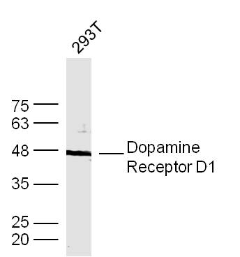
- Application Supplier NoteWB: 1:500-1:3000. ICC/IF: 1:100-1:1000. IHC-P: 1:100-1:1000. IHC-Fr: 1:100-1:1000. *Optimal dilutions/concentrations should be determined by the researcher.Not tested in other applications.
- ApplicationsImmunoFluorescence, Western Blot, ImmunoCytoChemistry, ImmunoHistoChemistry, ImmunoHistoChemistry Frozen, ImmunoHistoChemistry Paraffin
- CertificationResearch Use Only
- ClonalityPolyclonal
- Concentration2 mg/ml
- ConjugateUnconjugated
- Gene ID1812
- Target nameDRD1
- Target descriptiondopamine receptor D1
- Target synonymsD1R, DADR, DRD1A, D(1A) dopamine receptor, dopamine D1 receptor
- HostRabbit
- IsotypeIgG
- Protein IDP21728
- Protein NameD(1A) dopamine receptor
- Scientific DescriptionThis gene encodes the D1 subtype of the dopamine receptor. The D1 subtype is the most abundant dopamine receptor in the central nervous system. This G-protein coupled receptor stimulates adenylyl cyclase and activates cyclic AMP-dependent protein kinases. D1 receptors regulate neuronal growth and development, mediate some behavioral responses, and modulate dopamine receptor D2-mediated events. Alternate transcription initiation sites result in two transcript variants of this gene. [provided by RefSeq]
- ReactivityHuman, Mouse, Rat
- Storage Instruction-20°C or -80°C,2°C to 8°C
- UNSPSC41116161
Datasheet
Related products
Product group Antibodies
Anti-DRD1 AntibodyA96213
ApplicationsImmunoFluorescence, ELISA, ImmunoHistoChemistry
ReactivityHuman, Mouse, Rat
- SizePrice
Product group Antibodies
Anti-DRD1 Antibody144-02893
ApplicationsWestern Blot
ReactivityHuman, Mouse, Rat
TargetDRD1
- SizePrice
Product group Antibodies
References
DRD1 Polyclonal AntibodyBS-1007R
ApplicationsFlow Cytometry, ImmunoFluorescence, ImmunoHistoChemistry, ImmunoHistoChemistry Frozen, ImmunoHistoChemistry Paraffin
ReactivityHuman, Mouse, Rat
TargetDRD1
- SizePrice
Product group Antibodies
DRD1 AntibodyCSB-PA007178LA01HU
ApplicationsImmunoFluorescence, Western Blot, ELISA, ImmunoHistoChemistry
ReactivityHuman
TargetDRD1
- SizePrice
Product group Antibodies
DRD1 Polyclonal AntibodyCAC14639
ApplicationsImmunoFluorescence, Western Blot, ELISA, ImmunoHistoChemistry
TargetDRD1
- SizePrice

Product group Antibodies
ApplicationsWestern Blot
ReactivityHuman, Mouse, Rat
TargetDRD1
- SizePrice
Product group Antibodies

DRD1 / Dopamine Receptor D1 AntibodyLS-C402231
ApplicationsWestern Blot, ELISA, ImmunoHistoChemistry
ReactivityHuman
TargetDRD1
- SizePrice
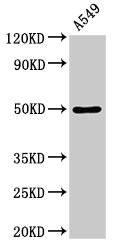
![Mouse tissue extract (30 μg) was separated by 10% SDS-PAGE, and the membrane was blotted with Dopamine Receptor D1 antibody [HL2553] (GTX638925) diluted at 1:500. The HRP-conjugated anti-rabbit IgG antibody (GTX213110-01) was used to detect the primary antibody.](https://www.genetex.com/upload/website/prouct_img/normal/GTX638925/GTX638925_45313_20250124_WB_M_brain_25020422_962.webp)
Product group Antibodies
ApplicationsFlow Cytometry, Western Blot
ReactivityHuman, Mouse
TargetDRD1
- SizePrice
![Non-transfected (–) and transfected (+) unboiled 293T whole cell extracts were separated by 10% SDS-PAGE, and the membrane was blotted with Dopamine Receptor D1 antibody [HL2680] (GTX639344) diluted at 1:5000. The HRP-conjugated anti-rabbit IgG antibody (GTX213110-01) was used to detect the primary antibody.](https://www.genetex.com/upload/website/prouct_img/normal/GTX639344/GTX639344_T-45222_20240216_WB_multiple_B_24021917_587.webp)
Product group Antibodies
ApplicationsWestern Blot
ReactivityHuman
TargetDRD1
- SizePrice