

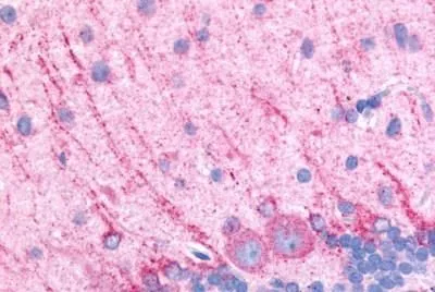
IHC-P analysis of rat brain, purkinje neurons tissue using GTX77969 Dopamine Receptor D5 antibody. Antigen retrieval : Heat-induced antigen retrieval
IHC-P analysis of rat brain, purkinje neurons tissue using GTX77969 Dopamine Receptor D5 antibody. Antigen retrieval : Heat-induced antigen retrieval
Dopamine Receptor D5 antibody
GTX77969
ApplicationsWestern Blot, ImmunoHistoChemistry, ImmunoHistoChemistry Frozen, ImmunoHistoChemistry Paraffin
Product group Antibodies
ReactivityHuman, Mouse, Rat, Other Species
TargetDRD5
Overview
- SupplierGeneTex
- Product NameDopamine Receptor D5 antibody
- Delivery Days Customer9
- Application Supplier NoteIHC-P: 3.4 microg/ml. IHC-P: 3.4 microg/ml. *Optimal dilutions/concentrations should be determined by the researcher.Not tested in other applications.
- ApplicationsWestern Blot, ImmunoHistoChemistry, ImmunoHistoChemistry Frozen, ImmunoHistoChemistry Paraffin
- CertificationResearch Use Only
- ClonalityPolyclonal
- Concentration1 mg/ml
- ConjugateUnconjugated
- Gene ID1816
- Target nameDRD5
- Target descriptiondopamine receptor D5
- Target synonymsDBDR, DRD1B, DRD1L2, D(1B) dopamine receptor, D1beta dopamine receptor, d(5) dopamine receptor, dopamine D5 receptor, dopamine receptor D1B
- HostRabbit
- IsotypeIgG
- Protein IDP21918
- Protein NameD(1B) dopamine receptor
- Scientific DescriptionThis gene encodes the D5 subtype of the dopamine receptor. The D5 subtype is a G-protein coupled receptor which stimulates adenylyl cyclase. This receptor is expressed in neurons in the limbic regions of the brain. It has a 10-fold higher affinity for dopamine than the D1 subtype. Pseudogenes related to this gene reside on chromosomes 1 and 2. [provided by RefSeq, Jul 2008]
- ReactivityHuman, Mouse, Rat, Other Species
- Storage Instruction-20°C or -80°C,2°C to 8°C
- UNSPSC41116161
Datasheet
Related products
Product group Antibodies
Anti-DRD5 AntibodyA285965
ApplicationsFlow Cytometry, ImmunoFluorescence, ELISA
ReactivityHuman
- SizePrice

Product group Antibodies
Anti-DRD5 Antibody Picoband(r)A03010-1-CARRIER-FREE
ApplicationsFlow Cytometry, Western Blot, ELISA
ReactivityHuman, Mouse, Rat
TargetDRD5
- SizePrice
Product group Antibodies
Anti-PDP2 Antibody144-64831
ApplicationsImmunoFluorescence, Western Blot
ReactivityHuman, Mouse, Rat
TargetDRD5
- SizePrice
Product group Antibodies
DRD5 AntibodyCSB-PA007182EA01HU
ApplicationsImmunoFluorescence, Western Blot, ELISA, ImmunoHistoChemistry
ReactivityHuman, Mouse
TargetDRD5
- SizePrice
Product group Antibodies

Goat anti-DRD5EB07424
ApplicationsFlow Cytometry, ImmunoFluorescence, ELISA
ReactivityHuman
TargetDRD5
- SizePrice
Product group Antibodies
Drd5 Polyclonal AntibodyCAC07349
ApplicationsImmunoFluorescence, Western Blot, ELISA, ImmunoHistoChemistry
ReactivityMouse
TargetDRD5
- SizePrice
Product group Antibodies

DRD5 / Dopamine Receptor D5 AntibodyLS-C400700
ApplicationsWestern Blot, ELISA, ImmunoHistoChemistry
ReactivityHuman
TargetDRD5
- SizePrice
![Dopamine Receptor D5 antibody [HL3081] detects Dopamine Receptor D5 protein by immunofluorescent analysis. Sample: SH-SY-5Y cells were fixed in ice-cold MeOH for 5 min. Green: Dopamine Receptor D5 stained by Dopamine Receptor D5 antibody [HL3081] (GTX640527) diluted at 1:500. Red: alpha Tubulin, a cytoskeleton marker, stained by alpha Tubulin antibody [GT114] (GTX628802) diluted at 1:1000. Blue: Fluoroshield with DAPI (GTX30920).](https://www.genetex.com/upload/website/prouct_img/normal/GTX640527/GTX640527_T-45446_20240712_ICC_IF_24080622_450.webp)
Product group Antibodies
ApplicationsImmunoFluorescence, ImmunoCytoChemistry, ImmunoHistoChemistry, ImmunoHistoChemistry Paraffin
ReactivityHuman, Mouse
TargetDRD5
- SizePrice
Product group Antibodies
Anti-PDP2 AntibodyCAB17190
ApplicationsImmunoFluorescence, Western Blot, ELISA, ImmunoCytoChemistry
ReactivityMouse
TargetDRD5
- SizePrice