

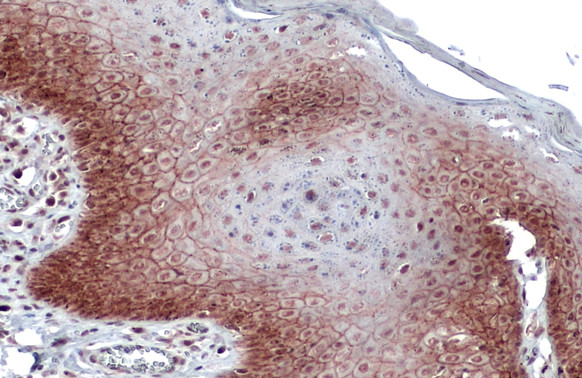
E-Cadherin antibody [GT477] detects E-Cadherin protein at cell membrane and cytoplasm by immunohistochemical analysis. Sample: Paraffin-embedded dog skin. E-Cadherin stained by E-Cadherin antibody [GT477] (GTX629691) diluted at 1:200. Antigen Retrieval: Citrate buffer, pH 6.0, 15 min
![Whole cell extract (30 μg) was separated by 7.5% SDS-PAGE, and the membrane was blotted with E-Cadherin antibody [GT477] (GTX629691) diluted at 1:2000. The HRP-conjugated anti-mouse IgG antibody (GTX213111-01) was used to detect the primary antibody.](https://www.genetex.com/upload/website/prouct_img/normal/GTX629691/GTX629691_41400_20181123_WB_D_22111423_113.webp "Whole cell extract (30 μg) was separated by 7.5% SDS-PAGE, and the membrane was blotted with E-Cadherin antibody [GT477] (GTX629691) diluted at 1:2000. The HRP-conjugated anti-mouse IgG antibody (GTX213111-01) was used to detect the primary antibody.")
![E-Cadherin antibody [GT477] detects E-Cadherin protein at cell membrane by immunofluorescent analysis. Sample: MDCK cells were fixed in 4% paraformaldehyde at RT for 15 min. Green: E-Cadherin stained by E-Cadherin antibody [GT477] (GTX629691) diluted at 1:2000.](https://www.genetex.com/upload/website/prouct_img/normal/GTX629691/GTX629691_41400_20181206_ICC_IF_D_22111423_770.webp "E-Cadherin antibody [GT477] detects E-Cadherin protein at cell membrane by immunofluorescent analysis. Sample: MDCK cells were fixed in 4% paraformaldehyde at RT for 15 min. Green: E-Cadherin stained by E-Cadherin antibody [GT477] (GTX629691) diluted at 1:2000.")
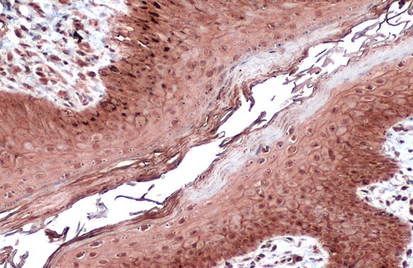
![E-Cadherin antibody [GT477] detects E-Cadherin protein at cell membrane and cytoplasm in mouse intestine by immunohistochemical analysis. Sample: Paraffin-embedded mouse intestine. E-Cadherin antibody [GT477] (GTX629691) diluted at 1:250.
Antigen Retrieval: Citrate buffer, pH 6.0, 15 min](https://www.genetex.com/upload/website/prouct_img/normal/GTX629691/GTX629691_41400_20160816_IHC-P_M_w_23061202_655.webp "E-Cadherin antibody [GT477] detects E-Cadherin protein at cell membrane and cytoplasm in mouse intestine by immunohistochemical analysis. Sample: Paraffin-embedded mouse intestine. E-Cadherin antibody [GT477] (GTX629691) diluted at 1:250.
Antigen Retrieval: Citrate buffer, pH 6.0, 15 min")
![Whole cell extract (30 μg) was separated by 5% SDS-PAGE, and the membrane was blotted with E-Cadherin antibody [GT477] (GTX629691) diluted at 1:500. The HRP-conjugated anti-mouse IgG antibody (GTX213111-01) was used to detect the primary antibody.](https://www.genetex.com/upload/website/prouct_img/normal/GTX629691/GTX629691_41400_20210416_WB_R_w_23061202_262.webp "Whole cell extract (30 μg) was separated by 5% SDS-PAGE, and the membrane was blotted with E-Cadherin antibody [GT477] (GTX629691) diluted at 1:500. The HRP-conjugated anti-mouse IgG antibody (GTX213111-01) was used to detect the primary antibody.")
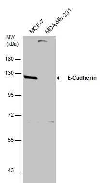
![E-Cadherin antibody [GT477] (GTX629691) detects E-Cadherin protein by flow cytometry analysis. Sample: MCF-7 cell. Black: Unlabelled sample was used as a control. Red: E-Cadherin antibody [GT477] (GTX629691) dilution: 1:50. Acquisition of 20,000 events were collected for FACS analysis.](https://www.genetex.com/upload/website/prouct_img/normal/GTX629691/GTX629691_41400_20181207_FACS_w_23061202_811.webp "E-Cadherin antibody [GT477] (GTX629691) detects E-Cadherin protein by flow cytometry analysis. Sample: MCF-7 cell. Black: Unlabelled sample was used as a control. Red: E-Cadherin antibody [GT477] (GTX629691) dilution: 1:50. Acquisition of 20,000 events were collected for FACS analysis.")
![Various whole cell extracts (30 μg) were separated by 5% SDS-PAGE, and the membrane was blotted with E-Cadherin antibody [GT477] (GTX629691) diluted at 1:3000. The HRP-conjugated anti-mouse IgG antibody (GTX213111-01) was used to detect the primary antibody.](https://www.genetex.com/upload/website/prouct_img/normal/GTX629691/GTX629691_44552_20220107_WB_w_23061202_393.webp "Various whole cell extracts (30 μg) were separated by 5% SDS-PAGE, and the membrane was blotted with E-Cadherin antibody [GT477] (GTX629691) diluted at 1:3000. The HRP-conjugated anti-mouse IgG antibody (GTX213111-01) was used to detect the primary antibody.")
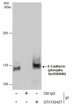
![E-adherin antibody [GT477] immunoprecipitates E-adherin protein in IP experiments. IP samples: MCF-7 whole cell extract A. Control with 3 μg of preimmune Mouse IgG B. Immunoprecipitation of E-adherin protein by 3 μg E-adherin antibody [GT477] (GTX629691) 5 % SDS-PAGE The immunoprecipitated E-adherin protein was detected by E-adherin antibody [GT477] (GTX629691) diluted at 1:500. [EasyBlot anti-mouse IgG (GTX221667-01) was used as a secondary reagent]](https://www.genetex.com/upload/website/prouct_img/normal/GTX629691/GTX629691_41400_IP_w_23061202_281.webp "E-adherin antibody [GT477] immunoprecipitates E-adherin protein in IP experiments. IP samples: MCF-7 whole cell extract A. Control with 3 μg of preimmune Mouse IgG B. Immunoprecipitation of E-adherin protein by 3 μg E-adherin antibody [GT477] (GTX629691) 5 % SDS-PAGE The immunoprecipitated E-adherin protein was detected by E-adherin antibody [GT477] (GTX629691) diluted at 1:500. [EasyBlot anti-mouse IgG (GTX221667-01) was used as a secondary reagent]")
![E-Cadherin antibody [GT477] detects E-Cadherin protein at cell membrane and cytoplasm in rat intestine by immunohistochemical analysis. Sample: Paraffin-embedded rat intestine. E-Cadherin antibody [GT477] (GTX629691) diluted at 1:250.
Antigen Retrieval: Citrate buffer, pH 6.0, 15 min](https://www.genetex.com/upload/website/prouct_img/normal/GTX629691/GTX629691_41400_20160816_IHC-P_R_w_23061202_382.webp "E-Cadherin antibody [GT477] detects E-Cadherin protein at cell membrane and cytoplasm in rat intestine by immunohistochemical analysis. Sample: Paraffin-embedded rat intestine. E-Cadherin antibody [GT477] (GTX629691) diluted at 1:250.
Antigen Retrieval: Citrate buffer, pH 6.0, 15 min")
![Mouse tissue extract (50 μg) was separated by 5% SDS-PAGE, and the membrane was blotted with E-Cadherin antibody [GT477] (GTX635000) diluted at 1:500. The HRP-conjugated anti-mouse IgG antibody (GTX213111-01) was used to detect the primary antibody.](https://www.genetex.com/upload/website/prouct_img/normal/GTX629691/GTX629691_41400_20210423_WB_M_intestine_w_23061202_656.webp "Mouse tissue extract (50 μg) was separated by 5% SDS-PAGE, and the membrane was blotted with E-Cadherin antibody [GT477] (GTX635000) diluted at 1:500. The HRP-conjugated anti-mouse IgG antibody (GTX213111-01) was used to detect the primary antibody.")
E-Cadherin antibody [GT477] detects E-Cadherin protein at cell membrane and cytoplasm by immunohistochemical analysis. Sample: Paraffin-embedded dog skin. E-Cadherin stained by E-Cadherin antibody [GT477] (GTX629691) diluted at 1:200. Antigen Retrieval: Citrate buffer, pH 6.0, 15 min
E-Cadherin antibody [GT477]
GTX629691
ApplicationsFlow Cytometry, ImmunoFluorescence, ImmunoPrecipitation, Western Blot, ImmunoCytoChemistry, ImmunoHistoChemistry, ImmunoHistoChemistry Paraffin
Product group Antibodies
ReactivityCanine, Human, Mouse, Rat
TargetCDH1
Overview
- SupplierGeneTex
- Product NameE-Cadherin antibody [GT477]
- Delivery Days Customer9
- Application Supplier NoteWB: 1:500-1:3000. ICC/IF: 1:100-1:1000. IHC-P: 1:100-1:1000. FACS: 1:50-1:200. IP: 1:100-1:500. *Optimal dilutions/concentrations should be determined by the researcher.Not tested in other applications.
- ApplicationsFlow Cytometry, ImmunoFluorescence, ImmunoPrecipitation, Western Blot, ImmunoCytoChemistry, ImmunoHistoChemistry, ImmunoHistoChemistry Paraffin
- CertificationResearch Use Only
- ClonalityMonoclonal
- Clone IDGT477
- Concentration1 mg/ml
- ConjugateUnconjugated
- Gene ID999
- Target nameCDH1
- Target descriptioncadherin 1
- Target synonymsArc-1, BCDS1, CD324, CDHE, ECAD, LCAM, UVO, cadherin-1, CAM 120/80, E-cadherin 1, cadherin 1, E-cadherin (epithelial), cadherin 1, type 1, E-cadherin (epithelial), calcium-dependent adhesion protein, epithelial, cell-CAM 120/80, epididymis secretory sperm binding protein, epithelial cadherin, uvomorulin
- HostMouse
- IsotypeIgG2a
- Protein IDP12830
- Protein NameCadherin-1
- Scientific DescriptionThis gene is a classical cadherin from the cadherin superfamily. The encoded protein is a calcium dependent cell-cell adhesion glycoprotein comprised of five extracellular cadherin repeats, a transmembrane region and a highly conserved cytoplasmic tail. Mutations in this gene are correlated with gastric, breast, colorectal, thyroid and ovarian cancer. Loss of function is thought to contribute to progression in cancer by increasing proliferation, invasion, and/or metastasis. The ectodomain of this protein mediates bacterial adhesion to mammalian cells and the cytoplasmic domain is required for internalization. Identified transcript variants arise from mutation at consensus splice sites. [provided by RefSeq]
- ReactivityCanine, Human, Mouse, Rat
- Storage Instruction-20°C or -80°C,2°C to 8°C
- UNSPSC12352203
References
- Adamiok-Ostrowska A, Grzanka M, Czarnocka B. Agrin is a novel oncogenic protein in thyroid cancer. Oncol Lett. 2023,26(5):483. doi: 10.3892/ol.2023.14070Read this paper
- Lima T, Barros AS, Trindade F, et al. Application of Proteogenomics to Urine Analysis towards the Identification of Novel Biomarkers of Prostate Cancer: An Exploratory Study. Cancers (Basel). 2022,14(8). doi: 10.3390/cancers14082001Read this paper
- Luo H, Yang Z, Zhang Q, et al. Carbon Ion Therapy Inhibits Esophageal Squamous Cell Carcinoma Metastasis by Upregulating STAT3 Through the JAK2/STAT3 Signaling Pathway. Front Public Health. 2020,8:579705. doi: 10.3389/fpubh.2020.579705Read this paper
- Pei YF, Xu XN, Wang ZF, et al. Methyl-CpG Binding Domain Protein 2 Inhibits the Malignant Characteristic of Lung Adenocarcinoma through the Epigenetic Modulation of 10 to 11 Translocation 1 and miR-200s. Am J Pathol. 2019,189(5):1065-1076. doi: 10.1016/j.ajpath.2019.01.010Read this paper
Datasheet
Related products
Product group Antibodies

Anti-E Cadherin [4A2]AB01700-1.1-BT
ApplicationsFlow Cytometry, ImmunoFluorescence, ImmunoPrecipitation, Western Blot, ImmunoHistoChemistry
ReactivityHuman, Mouse, Rat
TargetCDH1
- SizePrice
Product group Antibodies
Anti-CDH1 Antibody144-03044
ApplicationsImmunoFluorescence, Western Blot, ImmunoHistoChemistry
ReactivityHuman, Mouse, Rat
TargetCDH1
- SizePrice
Product group Antibodies
References
E-Cadherin antibody [N3C2], InternalGTX124178
ApplicationsImmunoFluorescence, ImmunoPrecipitation, Western Blot, ImmunoCytoChemistry, ImmunoHistoChemistry, ImmunoHistoChemistry Paraffin
ReactivityCanine, Human, Rat
TargetCDH1
- SizePrice
Product group Antibodies
References
E-Cadherin antibodyGTX124198
ApplicationsWestern Blot
ReactivityHuman
TargetCDH1
- SizePrice
Product group Antibodies
ApplicationsImmunoPrecipitation, Western Blot
ReactivityHuman, Mouse, Rat
TargetCDH1
- SizePrice
![FACS analysis of MCF-7 cells using GTX02618 E-Cadherin antibody [CDH1/2208R]. Blue : Primary antibody Red : Isotype control](https://www.genetex.com/upload/website/prouct_img/normal/GTX02618/GTX02618_20210319_FACS_w_23053122_361.webp)
Product group Antibodies
References
E-Cadherin antibody [CDH1/2208R]GTX02618
ApplicationsFlow Cytometry, ImmunoFluorescence, Western Blot, ImmunoCytoChemistry, ImmunoHistoChemistry, ImmunoHistoChemistry Paraffin
ReactivityHuman
TargetCDH1
- SizePrice
![IHC-P analysis of human invasive breast cancer of no special type (NST) tissue using GTX04367 E-Cadherin antibody [MSVA-035R] HistoMAX?. Strong membranous E-Cadherin staining in a breast cancer NST.](https://www.genetex.com/upload/website/prouct_img/normal/GTX04367/GTX04367_20230728_IHC-P_48_23072722_741.webp)
Product group Antibodies
ApplicationsImmunoHistoChemistry, ImmunoHistoChemistry Paraffin
ReactivityHuman
TargetCDH1
- SizePrice
![IHC-P analysis of human breast tissue using GTX57174 E-cadherin antibody [IHC564] (10X)](https://www.genetex.com/upload/website/prouct_img/normal/GTX57174/GTX57174_20180619_IHC-P_w_23061123_877.webp)
Product group Antibodies
E-Cadherin antibody [IHC564]GTX57174
ApplicationsImmunoHistoChemistry, ImmunoHistoChemistry Paraffin
ReactivityHuman
TargetCDH1
- SizePrice
![E-Cadherin antibody [GT311] (GTX629692) detects E-Cadherin protein by flow cytometry analysis. Sample: MCF-7 cell. Black: Unlabelled sample was used as a control. Red: E-Cadherin antibody [GT311] (GTX629692) dilution: 1:50. Acquisition of 20,000 events were collected for FACS analysis.](https://www.genetex.com/upload/website/prouct_img/normal/GTX629692/GTX629692_41400_20181207_FACS_w_23061202_932.webp)
Product group Antibodies
E-Cadherin antibody [GT311]GTX629692
ApplicationsFlow Cytometry, ImmunoPrecipitation, Western Blot
ReactivityHuman
TargetCDH1
- SizePrice