

E2EPF Antibody
ABX031529
ApplicationsWestern Blot, ELISA, ImmunoHistoChemistry
Product group Antibodies
Overview
- SupplierAbbexa
- Product NameE2EPF Antibody
- Delivery Days Customer12
- ApplicationsWestern Blot, ELISA, ImmunoHistoChemistry
- CertificationResearch Use Only
- ClonalityPolyclonal
- ConjugateUnconjugated
- HostRabbit
- UNSPSC12352203
Related products
Product group Antibodies
Anti-UBE2S AntibodyA285932
ApplicationsFlow Cytometry, ImmunoFluorescence, ELISA
ReactivityHuman
- SizePrice

Product group Antibodies
Anti-UBE2S Antibody Picoband(r)A03045-2-CARRIER-FREE
ApplicationsFlow Cytometry, ImmunoFluorescence, Western Blot, ELISA, ImmunoCytoChemistry
ReactivityHuman, Rat
TargetUBE2S
- SizePrice
Product group Antibodies
Anti-UBE2S Antibody144-04658
ApplicationsWestern Blot, ImmunoHistoChemistry
ReactivityHuman, Mouse, Rat
TargetUBE2S
- SizePrice
Product group Antibodies
E2EPF Polyclonal AntibodyBS-2305R
ApplicationsImmunoFluorescence, Western Blot, ELISA, ImmunoCytoChemistry, ImmunoHistoChemistry, ImmunoHistoChemistry Frozen, ImmunoHistoChemistry Paraffin
ReactivityBovine, Canine, Equine, Human, Mouse, Porcine, Rabbit, Rat
TargetUBE2S
- SizePrice
Product group Antibodies
UBE2S AntibodyCSB-PA142614
ApplicationsWestern Blot, ELISA, ImmunoHistoChemistry
ReactivityHuman, Mouse, Rat
TargetUBE2S
- SizePrice
Product group Antibodies

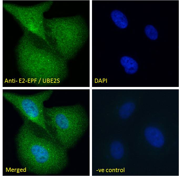
Goat anti-E2-EPF / UBE2SEB05758
ApplicationsFlow Cytometry, ImmunoFluorescence, ELISA
ReactivityBovine, Canine, Human, Mouse, Rat
TargetUBE2S
- SizePrice
Product group Antibodies
ApplicationsWestern Blot, ImmunoHistoChemistry
TargetUBE2S
- SizePrice
Product group Antibodies

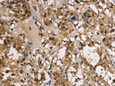
UBE2S / E2 EPF AntibodyLS-C404913
ApplicationsELISA, ImmunoHistoChemistry
ReactivityHuman, Mouse, Rat
TargetUBE2S
- SizePrice

Product group Antibodies
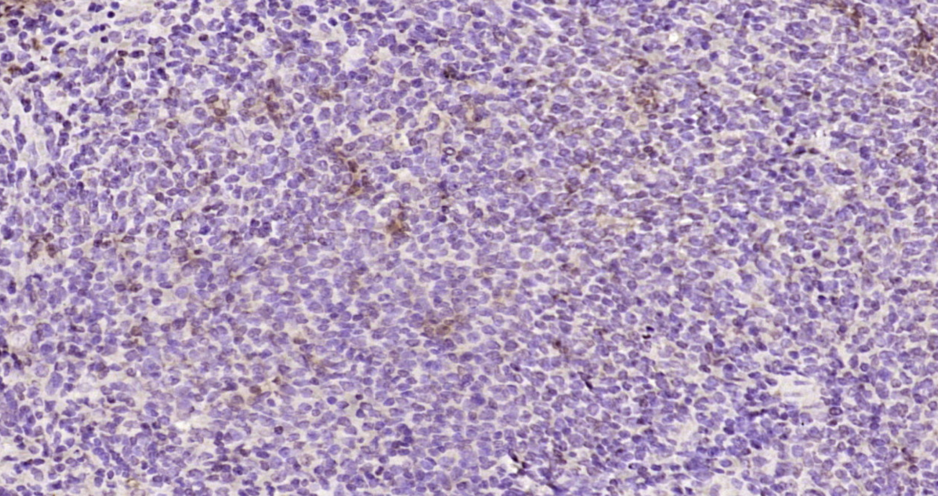
Anti-UBE2S AntibodyHPA057150
ApplicationsImmunoCytoChemistry, ImmunoHistoChemistry
ReactivityHuman
TargetUBE2S
- SizePrice
![Wild-type (WT) and UBE2S knockout (KO) 293T cell extracts (30 μg) were separated by 12% SDS-PAGE, and the membrane was blotted with UBE2S antibody [N1C2] (GTX115862) diluted at 1:2000. The HRP-conjugated anti-rabbit IgG antibody (GTX213110-01) was used to detect the primary antibody.](https://www.genetex.com/upload/website/prouct_img/normal/GTX115862/GTX115862_40296_20180105_WB_KO_watermark_w_23060519_223.webp)
Product group Antibodies
UBE2S antibody [N1C2]GTX115862
ApplicationsWestern Blot
ReactivityHuman
TargetUBE2S
- SizePrice