

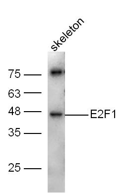
Mouse skeleton lysates probed with E2F1 Polyclonal Antibody, Unconjugated (bs-0599R) at 1:300 dilution and 4˚C overnight incubation. Followed by conjugated secondary antibody incubation at 1:10000 for 60 min at 37˚C.
, Unconjugated at 1:200, followed by conjugation to the secondary antibody and DAB staining")
at 1:50 dilution in blocking buffer and incubated for 30 min at room temperature, washed twice with 2%BSA in PBS, followed by secondary antibody incubation for 40 min at room temperature. Acquisitions of 20,000 events were performed. Cells stained with primary antibody (green), and isotype control (orange).")
Mouse skeleton lysates probed with E2F1 Polyclonal Antibody, Unconjugated (bs-0599R) at 1:300 dilution and 4˚C overnight incubation. Followed by conjugated secondary antibody incubation at 1:10000 for 60 min at 37˚C.
E2F1 Polyclonal Antibody
BS-0599R
ApplicationsFlow Cytometry, ImmunoFluorescence, Western Blot, ELISA, ImmunoCytoChemistry, ImmunoHistoChemistry, ImmunoHistoChemistry Frozen, ImmunoHistoChemistry Paraffin
Product group Antibodies
ReactivityBovine, Chicken, Equine, Human, Mouse, Porcine, Rabbit, Rat, Sheep
TargetE2F1
Overview
- SupplierBioss
- Product NameE2F1 Polyclonal Antibody
- Delivery Days Customer16
- ApplicationsFlow Cytometry, ImmunoFluorescence, Western Blot, ELISA, ImmunoCytoChemistry, ImmunoHistoChemistry, ImmunoHistoChemistry Frozen, ImmunoHistoChemistry Paraffin
- Applications SupplierWB(1:300-5000), ELISA(1:500-1000), FCM(1:20-100), IHC-P(1:200-400), IHC-F(1:100-500), IF(IHC-P)(1:50-200), IF(IHC-F)(1:50-200), IF(ICC)(1:50-200)
- CertificationResearch Use Only
- ClonalityPolyclonal
- Concentration1 ug/ul
- ConjugateUnconjugated
- Gene ID1869
- Target nameE2F1
- Target descriptionE2F transcription factor 1
- Target synonymsE2F-1, RBAP1, RBBP3, RBP3, transcription factor E2F1, PBR3, PRB-binding protein E2F-1, RBAP-1, RBBP-3, retinoblastoma-associated protein 1, retinoblastoma-binding protein 3, retinoblastoma-binding protein 3 (E2F-like)
- HostRabbit
- IsotypeIgG
- Protein IDQ01094
- Protein NameTranscription factor E2F1
- ReactivityBovine, Chicken, Equine, Human, Mouse, Porcine, Rabbit, Rat, Sheep
- Storage Instruction-20°C
- UNSPSC41116161
References
- Aloperine in combination with therapeutic adenoviral vector synergistically suppressed the growth of non-small cell lung cancer. Muhammad T et al., 2020 Apr, J Cancer Res Clin OncolRead this paper
- Silica nanoparticle exposure inducing granulosa cell apoptosis and follicular atresia in female Balb/c mice. Liu J et al., 2018 Feb, Environ Sci Pollut Res IntRead this paper
- Long-term PGC1beta overexpression leads to apoptosis, autophagy and muscle wasting. Sopariwala DH et al., 2017 Aug 31, Sci RepRead this paper
Datasheet
Related products
Product group Antibodies
Anti-E2F1 AntibodyA83673
ApplicationsImmunoFluorescence, ELISA
ReactivityDrosophila
- SizePrice
Product group Antibodies
Anti-E2F1 Antibody144-64611
ApplicationsImmunoFluorescence, Western Blot
ReactivityHuman, Mouse, Rat
TargetE2F1
- SizePrice
Product group Antibodies

Transcription factor E2F1 AntibodyABX433397
ApplicationsWestern Blot, ELISA
- SizePrice
Product group Antibodies

E2F1 AntibodyLS-C812948
ApplicationsWestern Blot, ELISA
ReactivityHuman, Rat
TargetE2F1
- SizePrice

Product group Antibodies
Anti-E2F1 Antibody Picoband(r)A00257-2-CARRIER-FREE
ApplicationsFlow Cytometry, Western Blot, ELISA
ReactivityHuman, Mouse, Rat
TargetE2F1
- SizePrice
Product group Antibodies
E2F1 AntibodyCSB-PA002235
ApplicationsWestern Blot, ELISA, ImmunoHistoChemistry
ReactivityHuman, Mouse
TargetE2F1
- SizePrice
Product group Antibodies

ApplicationsWestern Blot, ELISA
ReactivityHuman
TargetE2F1
- SizePrice
Product group Antibodies
E2F1 Polyclonal AntibodyCAC14530
ApplicationsImmunoPrecipitation, Western Blot, ELISA, ImmunoHistoChemistry
ReactivityMouse
TargetE2F1
- SizePrice
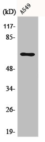
![Non-transfected (–) and transfected (+) 293T whole cell extracts (30 μg) were separated by 10% SDS-PAGE, and the membrane was blotted with E2F1 antibody [N1N3] (GTX101235) diluted at 1:500. The HRP-conjugated anti-rabbit IgG antibody (GTX213110-01) was used to detect the primary antibody.](https://www.genetex.com/upload/website/prouct_img/normal/GTX101235/GTX101235_39812_20181221_WB_B_w_23060100_187.webp)
Product group Antibodies
E2F1 antibody [N1N3]GTX101235
ApplicationsImmunoFluorescence, ImmunoPrecipitation, Western Blot, ImmunoCytoChemistry
ReactivityHuman
TargetE2F1
- SizePrice

Product group Antibodies
Anti-E2F1 AntibodyHPA029735
ApplicationsWestern Blot, ImmunoCytoChemistry
ReactivityHuman
TargetE2F1
- SizePrice