

EGF Receptor / EGFR Antibody
ORB750037
ApplicationsWestern Blot, ImmunoHistoChemistry, ImmunoHistoChemistry Paraffin
Product group Antibodies
ReactivityHuman
TargetEGFR
Overview
- SupplierBiorbyt
- Product NameEGF Receptor / EGFR Antibody
- Delivery Days Customer10
- Application Supplier NoteOptimal dilution of the EGF Receptor antibody should be determined by the researcher.
- ApplicationsWestern Blot, ImmunoHistoChemistry, ImmunoHistoChemistry Paraffin
- Applications SupplierImmunohistochemistry (FFPE): 0.1-0.2ug/ml for 30 min at RT,Western blot: 1-2ug/ml IHC-P, WB
- CertificationResearch Use Only
- ClonalityMonoclonal
- Clone IDGFR/1708
- ConjugateUnconjugated
- Gene ID1956
- Target nameEGFR
- Target descriptionepidermal growth factor receptor
- Target synonymsERBB, ERBB1, ERRP, HER1, NISBD2, NNCIS, PIG61, mENA, epidermal growth factor receptor, EGFR vIII, avian erythroblastic leukemia viral (v-erb-b) oncogene homolog, cell growth inhibiting protein 40, cell proliferation-inducing protein 61, epidermal growth factor receptor tyrosine kinase domain, erb-b2 receptor tyrosine kinase 1, proto-oncogene c-ErbB-1, receptor tyrosine-protein kinase erbB-1
- HostMouse
- IsotypeIgG1
- Protein IDP00533
- Protein NameEpidermal growth factor receptor
- Scientific DescriptionEpidermal growth factor receptor (EGFR) exists on the cell surface and is activated by binding of its specific ligands, including epidermal growth factor and transforming growth factor a. Upon activation by its growth factor ligands, EGFR undergoes a transition from an inactive monomeric form to an active homodimer. In addition to forming homodimers after ligand binding, EGFR may pair with another member of the ErbB receptor family, such as ErbB2/Her2/neu, to create an activated heterodimer. EGFR dimerization stimulates its intrinsic intracellular protein-tyrosine kinase activity. As a result, autophosphorylation of several tyrosine (Y) residues in the C-terminal domain of EGFR occurs. This autophosphorylation elicits downstream activation and signaling by several other proteins that associate with the phosphorylated tyrosines through their own phosphotyrosine-binding SH2 domains. These downstream signaling proteins initiate several signal transduction cascades, principally the MAPK, Akt and JNK pathways, leading to DNA synthesis and cell proliferation. [Wiki]
- ReactivityHuman
- Storage Instruction-20°C,2°C to 8°C
- UNSPSC12352203
Related products
Product group Antibodies

Anti-EGFR [C225 (Cetuximab)]Ab00279-1.1
ApplicationsFlow Cytometry, Neutralisation/Blocking
ReactivityHuman
TargetEGFR
- SizePrice
Product group Antibodies

EGFR-S1026 AntibodyABX032521
ApplicationsFlow Cytometry, Western Blot, ELISA, ImmunoHistoChemistry
- SizePrice
Product group Antibodies
ApplicationsFlow Cytometry
TargetEGFR
- SizePrice
Product group Antibodies
Anti-EGFR (N-term) Antibody131-0167
ApplicationsELISA
ReactivityHuman
TargetEGFR
- SizePrice

Product group Antibodies
Anti-EGFR Antibody Picoband(r)A00023-4-CARRIER-FREE
ApplicationsFlow Cytometry, ImmunoFluorescence, Western Blot, ELISA, ImmunoCytoChemistry, ImmunoHistoChemistry
ReactivityHuman, Mouse, Rat
TargetEGFR
- SizePrice
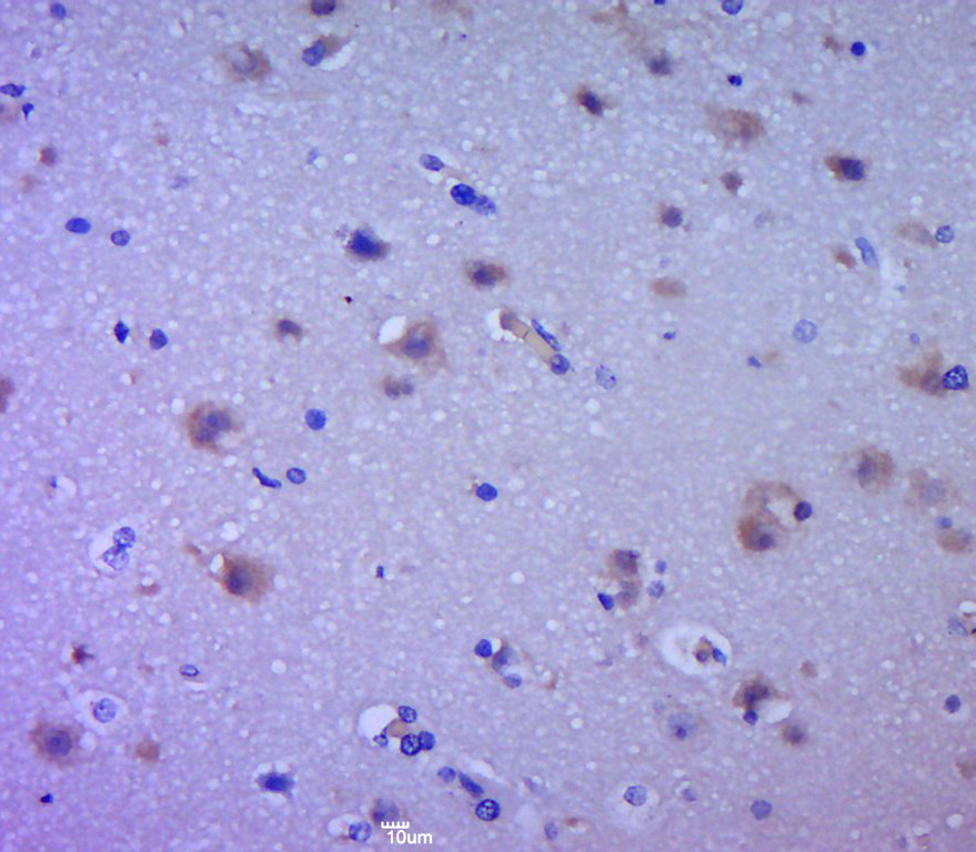
![EGFR antibody [C2C3], C-term detects EGFR protein at membrane on human gastric cancer by immunohistochemical analysis. Sample: Paraffin-embedded gastric cancer . EGFR antibody [C2C3], C-term (GTX100448) dilution: 1:500.
Antigen Retrieval: Trilogy? (EDTA based, pH 8.0) buffer, 15min](https://www.genetex.com/upload/website/prouct_img/normal/GTX100448/GTX100448_39645_IHC_w_23060100_679.webp)
Product group Antibodies
References
EGFR antibody [C2C3], C-termGTX100448
ApplicationsFlow Cytometry, ImmunoFluorescence, ImmunoPrecipitation, Western Blot, ImmunoCytoChemistry, ImmunoHistoChemistry, ImmunoHistoChemistry Paraffin
ReactivityHuman, Mouse, Rat
TargetEGFR
- SizePrice
Product group Antibodies
Anti-EGFRY058498
ApplicationsWestern Blot, ImmunoHistoChemistry
ReactivityHuman
- SizePrice
Product group Antibodies
Egfr Polyclonal AntibodyCAC09124
ApplicationsImmunoFluorescence, ELISA, ImmunoHistoChemistry
TargetEGFR
- SizePrice
Product group Antibodies
EGFR Polyclonal AntibodyBS-0165R
ApplicationsFlow Cytometry, ImmunoFluorescence, Western Blot, ELISA, ImmunoCytoChemistry, ImmunoHistoChemistry, ImmunoHistoChemistry Frozen, ImmunoHistoChemistry Paraffin
ReactivityCanine, Human, Mouse, Porcine, Rat
TargetEGFR
- SizePrice