

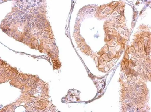
EGFR antibody [C2C3], C-term detects EGFR protein at membrane on human gastric cancer by immunohistochemical analysis. Sample: Paraffin-embedded gastric cancer . EGFR antibody [C2C3], C-term (GTX100448) dilution: 1:500.
Antigen Retrieval: Trilogy? (EDTA based, pH 8.0) buffer, 15min
![EGFR antibody [C2C3], C-term detects EGFR protein at membrane on human gastric cancer by immunohistochemical analysis. Sample: Paraffin-embedded gastric cancer. EGFR antibody [C2C3], C-term (GTX100448) dilution: 1:500.
Antigen Retrieval: Trilogy? (EDTA based, pH 8.0) buffer, 15min](https://www.genetex.com/upload/website/prouct_img/normal/GTX100448/GTX100448_39645_IHC_2_w_23060100_905.webp "EGFR antibody [C2C3], C-term detects EGFR protein at membrane on human gastric cancer by immunohistochemical analysis. Sample: Paraffin-embedded gastric cancer. EGFR antibody [C2C3], C-term (GTX100448) dilution: 1:500.
Antigen Retrieval: Trilogy? (EDTA based, pH 8.0) buffer, 15min")
![EGFR antibody [C2C3], C-term immunoprecipitates EGFR protein in IP experiments. IP samples: A431 whole cell extract A. 40 μg A431 whole cell extract B. Control with 4 μg of preimmune Rabbit IgG C. Immunoprecipitation of EGFR protein by 4 μg EGFR antibody [C2C3], C-term (GTX100448) 5 % SDS-PAGE The immunoprecipitated EGFR protein was detected by EGFR antibody [C2C3], C-term (GTX100448) diluted at 1:1000. [EasyBlot anti-rabbit IgG (GTX221666-01) was used as a secondary reagent]](https://www.genetex.com/upload/website/prouct_img/normal/GTX100448/GTX100448_39645_IP_w_23060100_264.webp "EGFR antibody [C2C3], C-term immunoprecipitates EGFR protein in IP experiments. IP samples: A431 whole cell extract A. 40 μg A431 whole cell extract B. Control with 4 μg of preimmune Rabbit IgG C. Immunoprecipitation of EGFR protein by 4 μg EGFR antibody [C2C3], C-term (GTX100448) 5 % SDS-PAGE The immunoprecipitated EGFR protein was detected by EGFR antibody [C2C3], C-term (GTX100448) diluted at 1:1000. [EasyBlot anti-rabbit IgG (GTX221666-01) was used as a secondary reagent]")
![Non-transfected (–) and transfected (+) A431 whole cell extracts (15 μg) were separated by 5% SDS-PAGE, and the membrane was blotted with EGFR antibody [C2C3], C-term (GTX100448) diluted at 1:6000. The HRP-conjugated anti-rabbit IgG antibody (GTX213110-01) was used to detect the primary antibody.](https://www.genetex.com/upload/website/prouct_img/normal/GTX100448/GTX100448_39645_20160630_WB_shRNA_watermark_w_23060100_225.webp "Non-transfected (–) and transfected (+) A431 whole cell extracts (15 μg) were separated by 5% SDS-PAGE, and the membrane was blotted with EGFR antibody [C2C3], C-term (GTX100448) diluted at 1:6000. The HRP-conjugated anti-rabbit IgG antibody (GTX213110-01) was used to detect the primary antibody.")
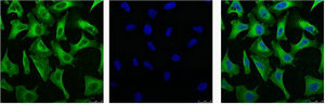
![EGFR antibody [C2C3], C-term detects EGFR protein at cell membrane by immunofluorescent analysis. Sample: A431 cells were fixed in ice-cold MeOH for 5 min. Green: EGFR stained by EGFR antibody [C2C3], C-term (GTX100448) diluted at 1:500. Blue: Fluoroshield with DAPI (GTX30920).](https://www.genetex.com/upload/website/prouct_img/normal/GTX100448/GTX100448_43782_20200806_ICC_IF_w_23060100_258.webp "EGFR antibody [C2C3], C-term detects EGFR protein at cell membrane by immunofluorescent analysis. Sample: A431 cells were fixed in ice-cold MeOH for 5 min. Green: EGFR stained by EGFR antibody [C2C3], C-term (GTX100448) diluted at 1:500. Blue: Fluoroshield with DAPI (GTX30920).")
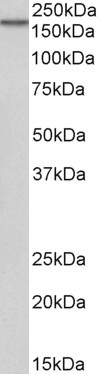
![Various whole cell extracts (30 μg) were separated by 5% SDS-PAGE, and the membrane was blotted with EGFR antibody [C2C3], C-term (GTX100448) diluted at 1:1000. The HRP-conjugated anti-rabbit IgG antibody (GTX213110-01) was used to detect the primary antibody.](https://www.genetex.com/upload/website/prouct_img/normal/GTX100448/GTX100448_43782_20191206_WB_w_23060100_713.webp "Various whole cell extracts (30 μg) were separated by 5% SDS-PAGE, and the membrane was blotted with EGFR antibody [C2C3], C-term (GTX100448) diluted at 1:1000. The HRP-conjugated anti-rabbit IgG antibody (GTX213110-01) was used to detect the primary antibody.")

![Various tissue extracts (50 μg) were separated by 5% SDS-PAGE, and the membrane was blotted with EGFR antibody [C2C3], C-term (GTX100448) diluted at 1:1000. The HRP-conjugated anti-rabbit IgG antibody (GTX213110-01) was used to detect the primary antibody. Corresponding RNA expression data are based on NCBI database.](https://www.genetex.com/upload/website/prouct_img/normal/GTX100448/GTX100448_45582_20250606_WB_M_tissue_RPKM_watermark_25061300_530.webp "Various tissue extracts (50 μg) were separated by 5% SDS-PAGE, and the membrane was blotted with EGFR antibody [C2C3], C-term (GTX100448) diluted at 1:1000. The HRP-conjugated anti-rabbit IgG antibody (GTX213110-01) was used to detect the primary antibody. Corresponding RNA expression data are based on NCBI database.")
EGFR antibody [C2C3], C-term detects EGFR protein at membrane on human gastric cancer by immunohistochemical analysis. Sample: Paraffin-embedded gastric cancer . EGFR antibody [C2C3], C-term (GTX100448) dilution: 1:500.
Antigen Retrieval: Trilogy? (EDTA based, pH 8.0) buffer, 15min
EGFR antibody [C2C3], C-term
GTX100448
ApplicationsFlow Cytometry, ImmunoFluorescence, ImmunoPrecipitation, Western Blot, ImmunoCytoChemistry, ImmunoHistoChemistry, ImmunoHistoChemistry Paraffin
Product group Antibodies
ReactivityHuman, Mouse, Rat
TargetEGFR
Overview
- SupplierGeneTex
- Product NameEGFR antibody [C2C3], C-term
- Delivery Days Customer9
- Application Supplier NoteWB: 1:500-1:3000. ICC/IF: 1:100-1:1000. IHC-P: 1:100-1:1000. IP: 1:100-1:500. *Optimal dilutions/concentrations should be determined by the researcher.Not tested in other applications.
- ApplicationsFlow Cytometry, ImmunoFluorescence, ImmunoPrecipitation, Western Blot, ImmunoCytoChemistry, ImmunoHistoChemistry, ImmunoHistoChemistry Paraffin
- CertificationResearch Use Only
- ClonalityPolyclonal
- Concentration0.28 mg/ml
- ConjugateUnconjugated
- Gene ID1956
- Target nameEGFR
- Target descriptionepidermal growth factor receptor
- Target synonymsERBB, ERBB1, ERRP, HER1, NISBD2, NNCIS, PIG61, mENA, epidermal growth factor receptor, EGFR vIII, avian erythroblastic leukemia viral (v-erb-b) oncogene homolog, cell growth inhibiting protein 40, cell proliferation-inducing protein 61, epidermal growth factor receptor tyrosine kinase domain, erb-b2 receptor tyrosine kinase 1, proto-oncogene c-ErbB-1, receptor tyrosine-protein kinase erbB-1
- HostRabbit
- IsotypeIgG
- Protein IDP00533
- Protein NameEpidermal growth factor receptor
- Scientific DescriptionThe protein encoded by this gene is a transmembrane glycoprotein that is a member of the protein kinase superfamily. This protein is a receptor for members of the epidermal growth factor family. EGFR is a cell surface protein that binds to epidermal growth factor. Binding of the protein to a ligand induces receptor dimerization and tyrosine autophosphorylation and leads to cell proliferation. Mutations in this gene are associated with lung cancer. [provided by RefSeq]
- ReactivityHuman, Mouse, Rat
- Storage Instruction-20°C or -80°C,2°C to 8°C
- UNSPSC41116161
Datasheet
Related products

Product group Antibodies
Anti-EGFR Antibody Picoband(r)A00023-4-CARRIER-FREE
ApplicationsFlow Cytometry, ImmunoFluorescence, Western Blot, ELISA, ImmunoCytoChemistry, ImmunoHistoChemistry
ReactivityHuman, Mouse, Rat
TargetEGFR
- SizePrice
Product group Antibodies
Anti-EGFR AntibodyA83204
ApplicationsWestern Blot, ELISA
ReactivityHuman
- SizePrice
Product group Antibodies

EGFR-S1026 AntibodyABX032521
ApplicationsFlow Cytometry, Western Blot, ELISA, ImmunoHistoChemistry
- SizePrice

Product group Antibodies
Anti-EGFR AntibodyAMAB90816
ApplicationsWestern Blot, ImmunoHistoChemistry
ReactivityHuman
TargetEGFR
- SizePrice
Product group Antibodies

Anti-EGFR [C225 (Cetuximab)]Ab00279-1.1
ApplicationsFlow Cytometry, Neutralisation/Blocking
ReactivityHuman
TargetEGFR
- SizePrice
Product group Antibodies
References

Goat anti-EGFREB06962
ApplicationsWestern Blot, ELISA
ReactivityHuman
TargetEGFR
- SizePrice
Product group Antibodies
EGFR Monoclonal AntibodyCSB-MA000199
ApplicationsImmunoFluorescence, Western Blot, ELISA, ImmunoHistoChemistry
ReactivityHuman
TargetEGFR
- SizePrice
Product group Antibodies

EGFR Antibody (phospho-Ser1045)LS-C343346
ApplicationsWestern Blot, ELISA
ReactivityHuman
TargetEGFR
- SizePrice
Product group Antibodies
EGFR (phospho Tyr1068) antibodyGTX25644
ApplicationsImmunoFluorescence, Western Blot, ImmunoCytoChemistry, ImmunoHistoChemistry
ReactivityHuman, Mouse, Primate, Rat
TargetEGFR
- SizePrice