

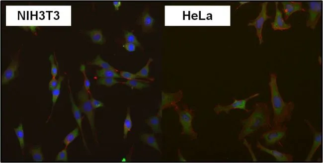
ICC/IF analysis of HeLa cells and NIH3T3 cells using GTX54748 eIF1 antibody [2B9]. Green : Primary antibody Blue : Nuclei Red : Actin Fixation : Formalin Permeabilization : 0.1% Triton X-100 in TBS for 10 minutes Dilution : 1:50 for at least 1 hour at room temperature
![ICC/IF analysis of MCF-7 cells using GTX54748 eIF1 antibody [2B9]. Cells were probed without (left) or with(right) an antibody. Green : Primary antibody Blue : Nuclei Red : Actin Fixation : Formalin Permeabilization : 0.1% Triton X-100 in TBS for 5-10 minute Dilution : 1:200 incubated overnight at 4 oC](https://www.genetex.com/upload/website/prouct_img/normal/GTX54748/GTX54748_802_ICC-IF_w_23060900_173.webp "ICC/IF analysis of MCF-7 cells using GTX54748 eIF1 antibody [2B9]. Cells were probed without (left) or with(right) an antibody. Green : Primary antibody Blue : Nuclei Red : Actin Fixation : Formalin Permeabilization : 0.1% Triton X-100 in TBS for 5-10 minute Dilution : 1:200 incubated overnight at 4 oC")
![WB analysis of 80μg whole cell lysate using GTX54748 eIF1 antibody [2B9]. Dilution : 1:1000](https://www.genetex.com/upload/website/prouct_img/normal/GTX54748/GTX54748_2129_WB_w_23060900_116.webp "WB analysis of 80μg whole cell lysate using GTX54748 eIF1 antibody [2B9]. Dilution : 1:1000")
![ICC/IF analysis of NIH-3T3 cells using GTX54748 eIF1 antibody [2B9]. Cells were probed without (left) or with(right) an antibody. Green : Primary antibody Blue : Nuclei Red : Actin Fixation : Formalin Permeabilization : 0.1% Triton X-100 in TBS for 5-10 minute Dilution : 1:200 incubated overnight at 4 oC](https://www.genetex.com/upload/website/prouct_img/normal/GTX54748/GTX54748_803_ICC-IF_w_23060900_309.webp "ICC/IF analysis of NIH-3T3 cells using GTX54748 eIF1 antibody [2B9]. Cells were probed without (left) or with(right) an antibody. Green : Primary antibody Blue : Nuclei Red : Actin Fixation : Formalin Permeabilization : 0.1% Triton X-100 in TBS for 5-10 minute Dilution : 1:200 incubated overnight at 4 oC")
![ICC/IF analysis of HeLa cells using GTX54748 eIF1 antibody [2B9]. Cells were probed without (left) or with(right) an antibody. Green : Primary antibody Blue : Nuclei Red : Actin Fixation : Formalin Permeabilization : 0.1% Triton X-100 in TBS for 5-10 minute Dilution : 1:200 incubated overnight at 4 oC](https://www.genetex.com/upload/website/prouct_img/normal/GTX54748/GTX54748_801_ICC-IF_w_23060900_785.webp "ICC/IF analysis of HeLa cells using GTX54748 eIF1 antibody [2B9]. Cells were probed without (left) or with(right) an antibody. Green : Primary antibody Blue : Nuclei Red : Actin Fixation : Formalin Permeabilization : 0.1% Triton X-100 in TBS for 5-10 minute Dilution : 1:200 incubated overnight at 4 oC")
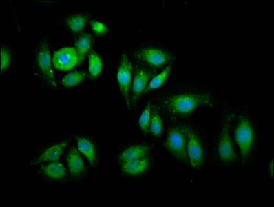
![ICC/IF analysis of MCF-7 cells using GTX54748 eIF1 antibody [2B9]. Panel e is a no primary antibody control. Green : Primary antibody Blue : Nuclei Red : Actin Fixation : 4% paraformaldehyde Permeabilization : 0.1% Triton? X-100 for 10 minutes Dilution : 2 μg/ml in 0.1% BSA and incubated for 3 hours at room temperature](https://www.genetex.com/upload/website/prouct_img/normal/GTX54748/GTX54748_800_ICC-IF_w_23060900_545.webp "ICC/IF analysis of MCF-7 cells using GTX54748 eIF1 antibody [2B9]. Panel e is a no primary antibody control. Green : Primary antibody Blue : Nuclei Red : Actin Fixation : 4% paraformaldehyde Permeabilization : 0.1% Triton? X-100 for 10 minutes Dilution : 2 μg/ml in 0.1% BSA and incubated for 3 hours at room temperature")
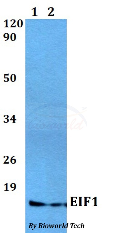
![WB analysis of whole cell extracts (30 μg lysate) of A549 (Lane 1), Hep G2 (lane 2) and PANC-1 (Lane 3) using GTX54748 eIF1 antibody [2B9]. Dilution : 1:500-1:2000](https://www.genetex.com/upload/website/prouct_img/normal/GTX54748/GTX54748_1887_WB_w_23060900_819.webp "WB analysis of whole cell extracts (30 μg lysate) of A549 (Lane 1), Hep G2 (lane 2) and PANC-1 (Lane 3) using GTX54748 eIF1 antibody [2B9]. Dilution : 1:500-1:2000")
ICC/IF analysis of HeLa cells and NIH3T3 cells using GTX54748 eIF1 antibody [2B9]. Green : Primary antibody Blue : Nuclei Red : Actin Fixation : Formalin Permeabilization : 0.1% Triton X-100 in TBS for 10 minutes Dilution : 1:50 for at least 1 hour at room temperature
eIF1 antibody [2B9]
GTX54748
ApplicationsImmunoFluorescence, Western Blot, ImmunoCytoChemistry
Product group Antibodies
ReactivityHuman, Mouse, Rat
TargetEIF1
Overview
- SupplierGeneTex
- Product NameeIF1 antibody [2B9]
- Delivery Days Customer9
- Application Supplier NoteWB: 1:500-1:2000. ICC/IF: 1-2 microg/ml. *Optimal dilutions/concentrations should be determined by the researcher.Not tested in other applications.
- ApplicationsImmunoFluorescence, Western Blot, ImmunoCytoChemistry
- CertificationResearch Use Only
- ClonalityMonoclonal
- Clone ID2B9
- Concentration1 mg/ml
- ConjugateUnconjugated
- Gene ID10209
- Target nameEIF1
- Target descriptioneukaryotic translation initiation factor 1
- Target synonymsA121, EIF-1, EIF1A, ISO1, SUI1, eukaryotic translation initiation factor 1, protein translation factor SUI1 homolog, sui1iso1
- HostMouse
- IsotypeIgG2a
- Protein IDP41567
- Protein NameEukaryotic translation initiation factor 1
- ReactivityHuman, Mouse, Rat
- Storage Instruction-20°C or -80°C,2°C to 8°C
- UNSPSC41116161
References
- DHX29 reduces leaky scanning through an upstream AUG codon regardless of its nucleotide context. Pisareva VP et al., 2016 May 19, Nucleic Acids ResRead this paper
Datasheet
Related products
Product group Antibodies
Anti-EIF1 AntibodyA28674
ApplicationsWestern Blot
ReactivityHuman, Mouse
- SizePrice
Product group Antibodies
Anti-EIF1 (C-term) Antibody102-24303
ApplicationsWestern Blot
TargetEIF1
- SizePrice

Product group Antibodies
Anti-EIF1 Antibody Picoband(r)A04125-1-CARRIER-FREE
ApplicationsFlow Cytometry, ImmunoFluorescence, Western Blot, ELISA, ImmunoCytoChemistry, ImmunoHistoChemistry
ReactivityHuman, Mouse, Rat
TargetEIF1
- SizePrice
Product group Antibodies
eIF1 Polyclonal AntibodyBS-13066R
ApplicationsImmunoFluorescence, Western Blot, ELISA, ImmunoCytoChemistry, ImmunoHistoChemistry, ImmunoHistoChemistry Frozen, ImmunoHistoChemistry Paraffin
ReactivityChicken, Equine, Human, Mouse, Porcine, Rat
TargetEIF1
- SizePrice
Product group Antibodies
EIF1 AntibodyCSB-PA007503LA01HU
ApplicationsImmunoFluorescence, ELISA
ReactivityHuman
TargetEIF1
- SizePrice
Product group Antibodies

EIF1 AntibodyLS-C497493
ApplicationsWestern Blot
ReactivityHuman, Mouse
TargetEIF1
- SizePrice