

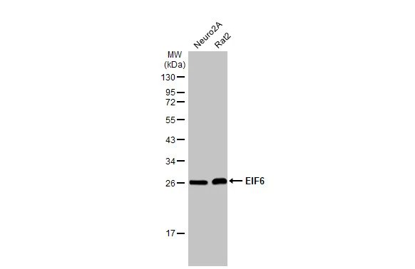
Various whole cell extracts (50 μg) were separated by 12% SDS-PAGE, and the membrane was blotted with EIF6 antibody [HL1758] (GTX637408) diluted at 1:1000. The HRP-conjugated anti-rabbit IgG antibody (GTX213110-01) was used to detect the primary antibody.
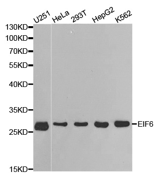
![Various whole cell extracts (30 μg) were separated by 12% SDS-PAGE, and the membrane was blotted with EIF6 antibody [HL1758] (GTX637408) diluted at 1:1000. The HRP-conjugated anti-rabbit IgG antibody (GTX213110-01) was used to detect the primary antibody.](https://www.genetex.com/upload/website/prouct_img/normal/GTX637408/GTX637408_44837_20221021_WB_22102723_194.webp "Various whole cell extracts (30 μg) were separated by 12% SDS-PAGE, and the membrane was blotted with EIF6 antibody [HL1758] (GTX637408) diluted at 1:1000. The HRP-conjugated anti-rabbit IgG antibody (GTX213110-01) was used to detect the primary antibody.")
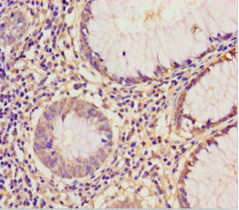
![EIF6 antibody [HL1758] detects EIF6 protein at cytoplasm and nucleus by immunohistochemical analysis. Sample: Paraffin-embedded human esophageal carcinoma. EIF6 stained by EIF6 antibody [HL1758] (GTX637408) diluted at 1:100. Antigen Retrieval: Citrate buffer, pH 6.0, 15 min](https://www.genetex.com/upload/website/prouct_img/normal/GTX637408/GTX637408_T-44788_20221007_IHC-P_22110201_320.webp "EIF6 antibody [HL1758] detects EIF6 protein at cytoplasm and nucleus by immunohistochemical analysis. Sample: Paraffin-embedded human esophageal carcinoma. EIF6 stained by EIF6 antibody [HL1758] (GTX637408) diluted at 1:100. Antigen Retrieval: Citrate buffer, pH 6.0, 15 min")
![Non-transfected (–) and transfected (+) 293T whole cell extracts (30 μg) were separated by 12% SDS-PAGE, and the membrane was blotted with EIF6 antibody [HL1758] (GTX637408) diluted at 1:5000. The HRP-conjugated anti-rabbit IgG antibody (GTX213110-01) was used to detect the primary antibody.](https://www.genetex.com/upload/website/prouct_img/normal/GTX637408/GTX637408_T-44788_20221118_WB_shRNA_watermark_22112219_885.webp "Non-transfected (–) and transfected (+) 293T whole cell extracts (30 μg) were separated by 12% SDS-PAGE, and the membrane was blotted with EIF6 antibody [HL1758] (GTX637408) diluted at 1:5000. The HRP-conjugated anti-rabbit IgG antibody (GTX213110-01) was used to detect the primary antibody.")
![Whole zebrafish extract (30 μg) was separated by 12% SDS-PAGE, and the membrane was blotted with EIF6 antibody [HL1758] (GTX637408) diluted at 1:1000. The HRP-conjugated anti-rabbit IgG antibody (GTX213110-01) was used to detect the primary antibody.](https://www.genetex.com/upload/website/prouct_img/normal/GTX637408/GTX637408_44837_20221216_WB_Z_22122018_668.webp "Whole zebrafish extract (30 μg) was separated by 12% SDS-PAGE, and the membrane was blotted with EIF6 antibody [HL1758] (GTX637408) diluted at 1:1000. The HRP-conjugated anti-rabbit IgG antibody (GTX213110-01) was used to detect the primary antibody.")
![Whole cell extract (30 μg) was separated by 12% SDS-PAGE, and the membrane was blotted with EIF6 antibody [HL1758] (GTX637408) diluted at 1:1000. The HRP-conjugated anti-rabbit IgG antibody (GTX213110-01) was used to detect the primary antibody.](https://www.genetex.com/upload/website/prouct_img/normal/GTX637408/GTX637408_44837_20230707_WB_Drosophila_23071223_990.webp "Whole cell extract (30 μg) was separated by 12% SDS-PAGE, and the membrane was blotted with EIF6 antibody [HL1758] (GTX637408) diluted at 1:1000. The HRP-conjugated anti-rabbit IgG antibody (GTX213110-01) was used to detect the primary antibody.")
![Whole Japanese medaka extract (30 μg) was separated by 12% SDS-PAGE, and the membrane was blotted with EIF6 antibody [HL1758] (GTX637408) diluted at 1:1000. The HRP-conjugated anti-rabbit IgG antibody (GTX213110-01) was used to detect the primary antibody, and the signal was developed with Trident ECL plus-Enhanced.](https://www.genetex.com/upload/website/prouct_img/normal/GTX637408/GTX637408_44837_20250815_WB_medaka_25082121_352.webp "Whole Japanese medaka extract (30 μg) was separated by 12% SDS-PAGE, and the membrane was blotted with EIF6 antibody [HL1758] (GTX637408) diluted at 1:1000. The HRP-conjugated anti-rabbit IgG antibody (GTX213110-01) was used to detect the primary antibody, and the signal was developed with Trident ECL plus-Enhanced.")
Various whole cell extracts (50 μg) were separated by 12% SDS-PAGE, and the membrane was blotted with EIF6 antibody [HL1758] (GTX637408) diluted at 1:1000. The HRP-conjugated anti-rabbit IgG antibody (GTX213110-01) was used to detect the primary antibody.
EIF6 antibody [HL1758]
GTX637408
ApplicationsWestern Blot, ImmunoHistoChemistry, ImmunoHistoChemistry Paraffin
Product group Antibodies
ReactivityDrosophila, Human, Mouse, Rat, Zebra Fish
TargetEIF6
Overview
- SupplierGeneTex
- Product NameEIF6 antibody [HL1758]
- Delivery Days Customer9
- Application Supplier NoteWB: 1:500-1:3000. *Optimal dilutions/concentrations should be determined by the researcher.Not tested in other applications.
- ApplicationsWestern Blot, ImmunoHistoChemistry, ImmunoHistoChemistry Paraffin
- CertificationResearch Use Only
- ClonalityMonoclonal
- Clone IDHL1758
- Concentration1 mg/ml
- ConjugateUnconjugated
- Gene ID3692
- Target nameEIF6
- Target descriptioneukaryotic translation initiation factor 6
- Target synonymsCAB, EIF3A, ITGB4BP, b(2)gcn, eIF-6, p27(BBP), p27BBP, eukaryotic translation initiation factor 6, B4 integrin interactor, eukaryotic translation initiation factor 3A, p27 beta-4 integrin-binding protein
- HostRabbit
- IsotypeIgG
- Protein IDP56537
- Protein NameEukaryotic translation initiation factor 6
- Scientific DescriptionHemidesmosomes are structures which link the basal lamina to the intermediate filament cytoskeleton. An important functional component of hemidesmosomes is the integrin beta-4 subunit (ITGB4), a protein containing two fibronectin type III domains. The protein encoded by this gene binds to the fibronectin type III domains of ITGB4 and may help link ITGB4 to the intermediate filament cytoskeleton. The encoded protein, which is insoluble and found both in the nucleus and in the cytoplasm, can function as a translation initiation factor and prevent the association of the 40S and 60S ribosomal subunits. Multiple non-protein coding transcript variants and variants encoding two different isoforms have been found for this gene. [provided by RefSeq, Jun 2012]
- ReactivityDrosophila, Human, Mouse, Rat, Zebra Fish
- Storage Instruction-20°C or -80°C,2°C to 8°C
- UNSPSC41116161
Datasheet
Related products
Product group Antibodies
Anti-EIF6 AntibodyA35495
ApplicationsImmunoFluorescence, Western Blot, ImmunoHistoChemistry
ReactivityHuman, Mouse, Rat
- SizePrice
Product group Antibodies
Anti-EIF6 Antibody144-01818
ApplicationsImmunoFluorescence, Western Blot
ReactivityHuman
TargetEIF6
- SizePrice
Product group Antibodies
eIF6 Polyclonal AntibodyBS-3844R
ApplicationsImmunoFluorescence, Western Blot, ELISA, ImmunoCytoChemistry, ImmunoHistoChemistry, ImmunoHistoChemistry Frozen, ImmunoHistoChemistry Paraffin
ReactivityBovine, Equine, Human, Mouse, Porcine, Rabbit, Rat, Sheep
TargetEIF6
- SizePrice
Product group Antibodies
EIF6 AntibodyCSB-PA007582LA01HU
ApplicationsImmunoFluorescence, ELISA, ImmunoHistoChemistry
ReactivityHuman
TargetEIF6
- SizePrice
Product group Antibodies
Eif6 Polyclonal AntibodyCAC11841
ApplicationsImmunoFluorescence, ELISA, ImmunoHistoChemistry
TargetEIF6
- SizePrice
Product group Antibodies

EIF6 AntibodyLS-C331705
ApplicationsImmunoFluorescence, Western Blot, ImmunoHistoChemistry
ReactivityHuman, Mouse, Rat
TargetEIF6
- SizePrice
Product group Antibodies
Anti-EIF6-25ulHPA040873
ApplicationsWestern Blot, ImmunoCytoChemistry, ImmunoHistoChemistry
ReactivityHuman, Mouse, Rat
- SizePrice
![Various tissue extracts (50 μg) were separated by 12% SDS-PAGE, and the membrane was blotted with EIF6 antibody [N1C3-2] (GTX117971) diluted at 1:5000. The HRP-conjugated anti-rabbit IgG antibody (GTX213110-01) was used to detect the primary antibody.](https://www.genetex.com/upload/website/prouct_img/normal/GTX117971/GTX117971_43649_20220923_WB_M_R_22092622_364.webp)
Product group Antibodies
EIF6 antibody [N1C3-2]GTX117971
ApplicationsWestern Blot, ImmunoHistoChemistry, ImmunoHistoChemistry Paraffin
ReactivityHuman, Mouse, Rat
TargetEIF6
- SizePrice
![Non-transfected (–) and transfected (+) 293T whole cell extracts (30 μg) were separated by 12% SDS-PAGE, and the membrane was blotted with EIF6 antibody [HL1759] (GTX637409) diluted at 1:5000. The HRP-conjugated anti-rabbit IgG antibody (GTX213110-01) was used to detect the primary antibody.](https://www.genetex.com/upload/website/prouct_img/normal/GTX637409/GTX637409_T-44788_20221118_WB_shRNA_watermark_22112219_525.webp)
Product group Antibodies
EIF6 antibody [HL1759]GTX637409
ApplicationsWestern Blot
ReactivityDrosophila, Human, Mouse, Rat
TargetEIF6
- SizePrice
Product group Antibodies
EIF6 antibodyGTX54010
ApplicationsImmunoFluorescence, Western Blot, ImmunoCytoChemistry
ReactivityHuman
TargetEIF6
- SizePrice