

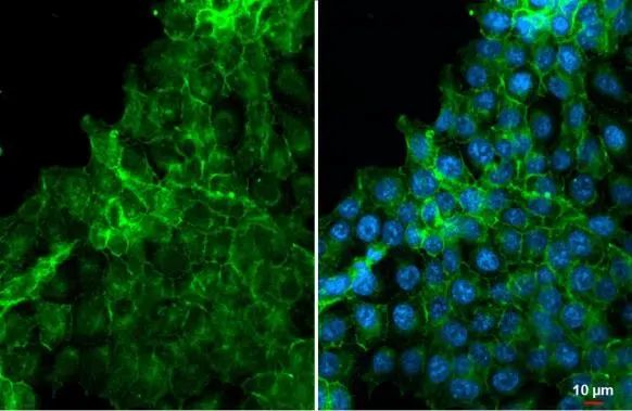
EpCAM antibody [HL1338] detects EpCAM protein at cell membrane and cell junction by immunofluorescent analysis. Sample: HCT116 cells were fixed in ice-cold MeOH for 5 min. Green: EpCAM stained by EpCAM antibody [HL1338] (GTX636758) diluted at 1:500. Blue: Fluoroshield with DAPI (GTX30920).
![EpCAM antibody [HL1338] (GTX636758) detects EpCAM protein by flow cytometry analysis. Sample: HCT-116 and HeLa cell. Gray: Rabbit IgG Isotype control. Red: EpCAM antibody [HL1338] (GTX636758) dilution: 1:50. Acquisition of 50,000 events were collected for flow cytometry analysis.](https://www.genetex.com/upload/website/prouct_img/normal/GTX636758/GTX636758_44662_20220829_FACS_22090701_882.webp "EpCAM antibody [HL1338] (GTX636758) detects EpCAM protein by flow cytometry analysis. Sample: HCT-116 and HeLa cell. Gray: Rabbit IgG Isotype control. Red: EpCAM antibody [HL1338] (GTX636758) dilution: 1:50. Acquisition of 50,000 events were collected for flow cytometry analysis.")
![EpCAM antibody [HL1338] detects EpCAM protein at cell membrane by immunohistochemical analysis. Sample: Paraffin-embedded mouse colon. EpCAM stained by EpCAM antibody [HL1338] (GTX636758) diluted at 1:100. Antigen Retrieval: Citrate buffer, pH 6.0, 15 min](https://www.genetex.com/upload/website/prouct_img/normal/GTX636758/GTX636758_T-44578_20220311_IHC-P_M_1_w_23061202_437.webp "EpCAM antibody [HL1338] detects EpCAM protein at cell membrane by immunohistochemical analysis. Sample: Paraffin-embedded mouse colon. EpCAM stained by EpCAM antibody [HL1338] (GTX636758) diluted at 1:100. Antigen Retrieval: Citrate buffer, pH 6.0, 15 min")
![EpCAM antibody [HL1338] detects EpCAM protein at cell membrane by immunohistochemical analysis. Sample: Paraffin-embedded mouse intestine. EpCAM stained by EpCAM antibody [HL1338] (GTX636758) diluted at 1:200. Antigen Retrieval: Citrate buffer, pH 6.0, 15 min](https://www.genetex.com/upload/website/prouct_img/normal/GTX636758/GTX636758_44662_20220527_IHC-P_M_w_23061202_112.webp "EpCAM antibody [HL1338] detects EpCAM protein at cell membrane by immunohistochemical analysis. Sample: Paraffin-embedded mouse intestine. EpCAM stained by EpCAM antibody [HL1338] (GTX636758) diluted at 1:200. Antigen Retrieval: Citrate buffer, pH 6.0, 15 min")
![Various whole cell extracts (30 μg) were separated by 10% SDS-PAGE, and the membrane was blotted with EpCAM antibody [HL1338] (GTX636758) diluted at 1:1000. The HRP-conjugated anti-rabbit IgG antibody (GTX213110-01) was used to detect the primary antibody.](https://www.genetex.com/upload/website/prouct_img/normal/GTX636758/GTX636758_44662_20220429_WB_w_23061202_751.webp "Various whole cell extracts (30 μg) were separated by 10% SDS-PAGE, and the membrane was blotted with EpCAM antibody [HL1338] (GTX636758) diluted at 1:1000. The HRP-conjugated anti-rabbit IgG antibody (GTX213110-01) was used to detect the primary antibody.")
![EpCAM antibody [HL1338] detects EpCAM protein at cell membrane by immunohistochemical analysis. Sample: Paraffin-embedded human HCT116 xenograft. EpCAM stained by EpCAM antibody [HL1338] (GTX636758) diluted at 1:200. Antigen Retrieval: Citrate buffer, pH 6.0, 15 min](https://www.genetex.com/upload/website/prouct_img/normal/GTX636758/GTX636758_44662_20220527_IHC-P_w_23061202_104.webp "EpCAM antibody [HL1338] detects EpCAM protein at cell membrane by immunohistochemical analysis. Sample: Paraffin-embedded human HCT116 xenograft. EpCAM stained by EpCAM antibody [HL1338] (GTX636758) diluted at 1:200. Antigen Retrieval: Citrate buffer, pH 6.0, 15 min")
EpCAM antibody [HL1338] detects EpCAM protein at cell membrane and cell junction by immunofluorescent analysis. Sample: HCT116 cells were fixed in ice-cold MeOH for 5 min. Green: EpCAM stained by EpCAM antibody [HL1338] (GTX636758) diluted at 1:500. Blue: Fluoroshield with DAPI (GTX30920).
EpCAM antibody [HL1338]
GTX636758
ApplicationsFlow Cytometry, ImmunoFluorescence, Western Blot, ImmunoCytoChemistry, ImmunoHistoChemistry, ImmunoHistoChemistry Paraffin
Product group Antibodies
ReactivityHuman, Mouse
TargetEPCAM
Overview
- SupplierGeneTex
- Product NameEpCAM antibody [HL1338]
- Delivery Days Customer9
- Application Supplier NoteIHC-P: 1:100-1:1000. *Optimal dilutions/concentrations should be determined by the researcher.Not tested in other applications.
- ApplicationsFlow Cytometry, ImmunoFluorescence, Western Blot, ImmunoCytoChemistry, ImmunoHistoChemistry, ImmunoHistoChemistry Paraffin
- CertificationResearch Use Only
- ClonalityMonoclonal
- Clone IDHL1338
- Concentration1 mg/ml
- ConjugateUnconjugated
- Gene ID4072
- Target nameEPCAM
- Target descriptionepithelial cell adhesion molecule
- Target synonymsBer-Ep4, BerEp4, DIAR5, EGP-2, EGP314, EGP40, ESA, HNPCC8, KS1/4, KSA, LYNCH8, M4S1, MIC18, MK-1, MOC-31, TACSTD1, TROP1, epithelial cell adhesion molecule, adenocarcinoma-associated antigen, cell surface glycoprotein Trop-1, epithelial glycoprotein 314, human epithelial glycoprotein-2, major gastrointestinal tumor-associated protein GA733-2, membrane component, chromosome 4, surface marker (35kD glycoprotein), trophoblast cell surface antigen 1, tumor-associated calcium signal transducer 1
- HostRabbit
- IsotypeIgG
- Protein IDP16422
- Protein NameEpithelial cell adhesion molecule
- Scientific DescriptionThis gene encodes a carcinoma-associated antigen and is a member of a family that includes at least two type I membrane proteins. This antigen is expressed on most normal epithelial cells and gastrointestinal carcinomas and functions as a homotypic calcium-independent cell adhesion molecule. The antigen is being used as a target for immunotherapy treatment of human carcinomas. Mutations in this gene result in congenital tufting enteropathy. [provided by RefSeq, Dec 2008]
- ReactivityHuman, Mouse
- Storage Instruction-20°C or -80°C,2°C to 8°C
- UNSPSC12352203
Datasheet
Related products
Product group Antibodies

Anti-EpCAM [AUA1]Ab00609-1.1
ApplicationsFlow Cytometry, ImmunoFluorescence, ELISA, ImmunoHistoChemistry
ReactivityHuman
TargetEPCAM
- SizePrice
Product group Antibodies
Anti-EPCAM Antibody144-01177
ApplicationsWestern Blot, ImmunoHistoChemistry
ReactivityHuman, Mouse
TargetEPCAM
- SizePrice

Product group Antibodies
Anti-EPCAM AntibodyAMAB91411
ApplicationsWestern Blot, ImmunoHistoChemistry
ReactivityHuman
TargetEPCAM
- SizePrice
![IHC-P analysis of human lung adenocarcinoma (LUAD) tissue using GTX04380 EpCAM antibody [MSVA-326R] HistoMAX?. Strong EpCAM staining in an adenocarcinoma of the lung.](https://www.genetex.com/upload/website/prouct_img/normal/GTX04380/GTX04380_20230728_IHC-P_50_23072722_768.webp)
Product group Antibodies
ApplicationsImmunoHistoChemistry, ImmunoHistoChemistry Paraffin
ReactivityHuman
TargetEPCAM
- SizePrice
![EpCAM antibody [N3C3] detects EpCAM protein at cell membrane and cell junction by immunofluorescent analysis. Sample: HCT116 cells were fixed in ice-cold MeOH for 5 min. Green: EpCAM stained by EpCAM antibody [N3C3] (GTX113091) diluted at 1:500. Blue: Fluoroshield with DAPI (GTX30920).](https://www.genetex.com/upload/website/prouct_img/normal/GTX113091/GTX113091_44265_20220624_ICC_IF_22062919_816.webp)
Product group Antibodies
References
EpCAM antibody [N3C3]GTX113091
ApplicationsImmunoFluorescence, Western Blot, ImmunoCytoChemistry, ImmunoHistoChemistry, ImmunoHistoChemistry Paraffin
ReactivityHuman, Mouse
TargetEPCAM
- SizePrice
![EpCAM antibody [GT7711] detects EpCAM protein at cell membrane by immunofluorescent analysis. Sample: HCT116 (left) and HeLa (right) cells were fixed in ice-cold MeOH for 5 min. Green: EpCAM stained by EpCAM antibody [GT7711] (GTX635473) diluted at 1:500. Blue: Fluoroshield with DAPI (GTX30920).](https://www.genetex.com/upload/website/prouct_img/normal/GTX635473/GTX635473_43913_20200722_ICC_IF_w_23051500_909.webp)
Product group Antibodies
EpCAM antibody [GT7711]GTX635473
ApplicationsImmunoFluorescence, Western Blot, ImmunoCytoChemistry
ReactivityHuman
TargetEPCAM
- SizePrice
![Various whole cell extracts (30 μg) were separated by 10% SDS-PAGE, and the membrane was blotted with EpCAM antibody [GT852] (GTX635474) diluted at 1:1000. The HRP-conjugated anti-mouse IgG antibody (GTX213111-01) was used to detect the primary antibody.](https://www.genetex.com/upload/website/prouct_img/normal/GTX635474/GTX635474_43913_20200515_WB_w_23061202_351.webp)
Product group Antibodies
EpCAM antibody [GT852]GTX635474
ApplicationsImmunoFluorescence, Western Blot, ImmunoCytoChemistry
ReactivityHuman
TargetEPCAM
- SizePrice
![Various whole cell extracts (30 μg) were separated by 10% SDS-PAGE, and the membranes were blotted with EpCAM antibody [GT25512] (GTX635967) diluted at 1:1000 and competitor's antibody diluted at 1:1000. The HRP-conjugated anti-mouse IgG antibody (GTX2131](https://www.genetex.com/upload/website/prouct_img/normal/GTX635967/GTX635967_44216_20210305_WB_competitor_watermark_w_23061202_505.webp)
Product group Antibodies
EpCAM antibody [GT25512]GTX635967
ApplicationsFlow Cytometry, ImmunoFluorescence, Western Blot, ImmunoCytoChemistry, ImmunoHistoChemistry, ImmunoHistoChemistry Paraffin
ReactivityHuman
TargetEPCAM
- SizePrice
![EpCAM antibody [GT3188] detects EpCAM protein at cell membrane by immunohistochemical analysis. Sample: Paraffin-embedded MDA-MB-231 xenograft. EpCAM stained by EpCAM antibody [GT3188] (GTX635970) diluted at 1:200. Antigen Retrieval: Citrate buffer, pH 6.0, 15 min](https://www.genetex.com/upload/website/prouct_img/normal/GTX635970/GTX635970_44216_20210326_IHC-P_w_23061202_241.webp)
Product group Antibodies
EpCAM antibody [GT3188]GTX635970
ApplicationsFlow Cytometry, ImmunoFluorescence, Western Blot, ImmunoCytoChemistry, ImmunoHistoChemistry, ImmunoHistoChemistry Paraffin
ReactivityHuman
TargetEPCAM
- SizePrice