

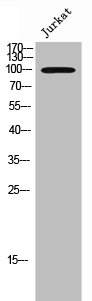
Whole cell extract (30 μg) was separated by 7.5% SDS-PAGE, and the membrane was blotted with EphA3 antibody [N1N3] (GTX114067) diluted at 1:1500. The HRP-conjugated anti-rabbit IgG antibody (GTX213110-01) was used to detect the primary antibody, and the signal was developed with Trident ECL plus-Enhanced.
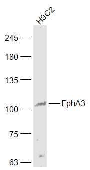
![Whole cell extract (30 μg) was separated by 7.5% SDS-PAGE, and the membrane was blotted with EphA3 antibody [N1N3] (GTX114067) diluted at 1:1500. The HRP-conjugated anti-rabbit IgG antibody (GTX213110-01) was used to detect the primary antibody.](https://www.genetex.com/upload/website/prouct_img/normal/GTX114067/GTX114067_40660_20211015_WB_w_23060501_972.webp "Whole cell extract (30 μg) was separated by 7.5% SDS-PAGE, and the membrane was blotted with EphA3 antibody [N1N3] (GTX114067) diluted at 1:1500. The HRP-conjugated anti-rabbit IgG antibody (GTX213110-01) was used to detect the primary antibody.")
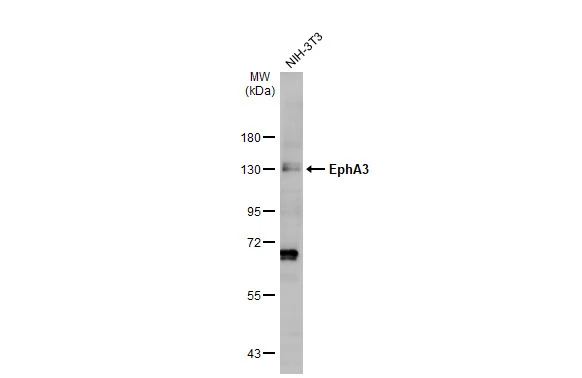
![Mouse tissue extract (50 μg) were separated by 7.5% SDS-PAGE, and the membrane was blotted with EphA3 antibody [N1N3] (GTX114067) diluted at 1:1500. The HRP-conjugated anti-rabbit IgG antibody (GTX213110-01) was used to detect the primary antibody.](https://www.genetex.com/upload/website/prouct_img/normal/GTX114067/GTX114067_40660_20211022_WB_M_tissue_w_23060501_807.webp "Mouse tissue extract (50 μg) were separated by 7.5% SDS-PAGE, and the membrane was blotted with EphA3 antibody [N1N3] (GTX114067) diluted at 1:1500. The HRP-conjugated anti-rabbit IgG antibody (GTX213110-01) was used to detect the primary antibody.")
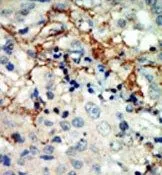
 antibody at 1:500 dilution.
Antigen Retrieval: Trilogy? (EDTA based, pH 8.0) buffer, 15min")
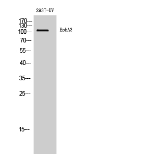
![Whole cell extract (30 μg) was separated by 7.5% SDS-PAGE, and the membrane was blotted with EphA3 antibody [N1N3] (GTX114067) diluted at 1:1500. The HRP-conjugated anti-rabbit IgG antibody (GTX213110-01) was used to detect the primary antibody.](https://www.genetex.com/upload/website/prouct_img/normal/GTX114067/GTX114067_44307_20210507_WB_w_23060501_260.webp "Whole cell extract (30 μg) was separated by 7.5% SDS-PAGE, and the membrane was blotted with EphA3 antibody [N1N3] (GTX114067) diluted at 1:1500. The HRP-conjugated anti-rabbit IgG antibody (GTX213110-01) was used to detect the primary antibody.")
![Various whole cell extracts (30 μg) were separated by 7.5% SDS-PAGE, and the membrane was blotted with EphA3 antibody [N1N3] (GTX114067) diluted at 1:1500. The HRP-conjugated anti-rabbit IgG antibody (GTX213110-01) was used to detect the primary antibody. Corresponding RNA expression data for the same cell lines are based on Human Protein Atlas program.](https://www.genetex.com/upload/website/prouct_img/normal/GTX114067/GTX114067_45299_20240906_WB_TPM_watermark_24091901_441.webp "Various whole cell extracts (30 μg) were separated by 7.5% SDS-PAGE, and the membrane was blotted with EphA3 antibody [N1N3] (GTX114067) diluted at 1:1500. The HRP-conjugated anti-rabbit IgG antibody (GTX213110-01) was used to detect the primary antibody. Corresponding RNA expression data for the same cell lines are based on Human Protein Atlas program.")
Whole cell extract (30 μg) was separated by 7.5% SDS-PAGE, and the membrane was blotted with EphA3 antibody [N1N3] (GTX114067) diluted at 1:1500. The HRP-conjugated anti-rabbit IgG antibody (GTX213110-01) was used to detect the primary antibody, and the signal was developed with Trident ECL plus-Enhanced.
EphA3 antibody [N1N3]
GTX114067
ApplicationsWestern Blot, ImmunoHistoChemistry, ImmunoHistoChemistry Paraffin
Product group Antibodies
ReactivityHuman, Mouse
TargetEPHA3
Overview
- SupplierGeneTex
- Product NameEphA3 antibody [N1N3]
- Delivery Days Customer9
- Application Supplier NoteWB: 1:500-1:3000. IHC-P: 1:100-1:1000. *Optimal dilutions/concentrations should be determined by the researcher.Not tested in other applications.
- ApplicationsWestern Blot, ImmunoHistoChemistry, ImmunoHistoChemistry Paraffin
- CertificationResearch Use Only
- ClonalityPolyclonal
- Concentration1 mg/ml
- ConjugateUnconjugated
- Gene ID2042
- Target nameEPHA3
- Target descriptionEPH receptor A3
- Target synonymsEK4, ETK, ETK1, HEK, HEK4, TYRO4, ephrin type-A receptor 3, EPH-like kinase 4, TYRO4 protein tyrosine kinase, eph-like tyrosine kinase 1, human embryo kinase 1, testicular tissue protein Li 64, tyrosine-protein kinase receptor ETK1
- HostRabbit
- IsotypeIgG
- Protein IDP29320
- Protein NameEphrin type-A receptor 3
- Scientific DescriptionThis gene belongs to the ephrin receptor subfamily of the protein-tyrosine kinase family. EPH and EPH-related receptors have been implicated in mediating developmental events, particularly in the nervous system. Receptors in the EPH subfamily typically have a single kinase domain and an extracellular region containing a Cys-rich domain and 2 fibronectin type III repeats. The ephrin receptors are divided into 2 groups based on the similarity of their extracellular domain sequences and their affinities for binding ephrin-A and ephrin-B ligands. This gene encodes a protein that binds ephrin-A ligands. Two alternatively spliced transcript variants have been described for this gene. [provided by RefSeq]
- ReactivityHuman, Mouse
- Storage Instruction-20°C or -80°C,2°C to 8°C
- UNSPSC41116161
Datasheet
Related products
Product group Antibodies
EPHA3 AntibodyCSB-PA008057
ApplicationsWestern Blot, ELISA
ReactivityHuman, Mouse, Rat
TargetEPHA3
- SizePrice
Product group Antibodies
Anti-EPHA3 AntibodyA97563
ApplicationsWestern Blot, ELISA
ReactivityHuman, Mouse, Rat
- SizePrice
Product group Antibodies

Anti-EphA3 [KB004]Ab02488-1.1
ApplicationsFunctional Assay
ReactivityHuman
TargetEPHA3
- SizePrice

Product group Antibodies
Anti-Eph receptor A3/EPHA3 Antibody Picoband(r)A02872-1-CARRIER-FREE
ApplicationsFlow Cytometry, ImmunoFluorescence, Western Blot, ELISA, ImmunoCytoChemistry, ImmunoHistoChemistry
ReactivityHuman
TargetEPHA3
- SizePrice
Product group Antibodies

EPHA3 / EPH Receptor A3 AntibodyLS-C831062
ApplicationsWestern Blot, ELISA
ReactivityHuman, Mouse, Rat
TargetEPHA3
- SizePrice

Product group Antibodies
Anti-EPHA3 AntibodyHPA069390
ApplicationsImmunoCytoChemistry
ReactivityHuman
TargetEPHA3
- SizePrice
Product group Antibodies
EphA3 Polyclonal AntibodyBS-7032R
ApplicationsImmunoFluorescence, Western Blot, ELISA, ImmunoCytoChemistry, ImmunoHistoChemistry, ImmunoHistoChemistry Frozen, ImmunoHistoChemistry Paraffin
ReactivityBovine, Canine, Equine, Human, Mouse, Rat, Sheep
TargetEPHA3
- SizePrice
![Various whole cell extracts (30 μg) were separated by 7.5% SDS-PAGE, and the membrane was blotted with EphA3 antibody [HL2651] (GTX639110) diluted at 1:1000. The HRP-conjugated anti-rabbit IgG antibody (GTX213110-01) was used to detect the primary antibody. Corresponding RNA expression data for the same cell lines are based on Human Protein Atlas program.](https://www.genetex.com/upload/website/prouct_img/normal/GTX639110/GTX639110_45299_20240126_WB_TPM_watermark_24013018_394.webp)
Product group Antibodies
EphA3 antibody [HL2651]GTX639110
ApplicationsWestern Blot, ImmunoHistoChemistry, ImmunoHistoChemistry Paraffin
ReactivityHuman, Mouse, Rat
TargetEPHA3
- SizePrice
Product group Antibodies
EphA3 antibody, N-termGTX81361
ApplicationsWestern Blot, ImmunoHistoChemistry, ImmunoHistoChemistry Paraffin
ReactivityHuman
TargetEPHA3
- SizePrice