

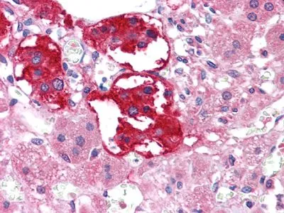
IHC-P analysis of human Adrenal tissue using GTX83041 EphB3 antibody [4A122D1].
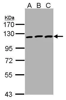
![WB analysis of truncated EphB3-His recombinant protein using GTX83041 EphB3 antibody [4A122D1].](https://www.genetex.com/upload/website/prouct_img/normal/GTX83041/GTX83041_20170912_WB_w_23061322_539.webp "WB analysis of truncated EphB3-His recombinant protein using GTX83041 EphB3 antibody [4A122D1].")
![IHC-P analysis of human lung squamous cell carcinoma (A), lung adenocarcinoma (B), colon carcinoma (C), breast carcinoma (D), normal sublingual gland (E), normal rectal (F) using GTX83041 EphB3 antibody [4A122D1].](https://www.genetex.com/upload/website/prouct_img/normal/GTX83041/GTX83041_20170912_IHC-P_1_w_23061322_445.webp "IHC-P analysis of human lung squamous cell carcinoma (A), lung adenocarcinoma (B), colon carcinoma (C), breast carcinoma (D), normal sublingual gland (E), normal rectal (F) using GTX83041 EphB3 antibody [4A122D1].")
IHC-P analysis of human Adrenal tissue using GTX83041 EphB3 antibody [4A122D1].
EphB3 antibody [4A122D1]
GTX83041
ApplicationsWestern Blot, ELISA, ImmunoHistoChemistry, ImmunoHistoChemistry Paraffin
Product group Antibodies
ReactivityHuman
TargetEPHB3
Overview
- SupplierGeneTex
- Product NameEphB3 antibody [4A122D1]
- Delivery Days Customer9
- Application Supplier NoteWB: 1/500 - 1/2000. IHC-P: 1/200 - 1/1000. ELISA: 1/10000. *Optimal dilutions/concentrations should be determined by the researcher.Not tested in other applications.
- ApplicationsWestern Blot, ELISA, ImmunoHistoChemistry, ImmunoHistoChemistry Paraffin
- CertificationResearch Use Only
- ClonalityMonoclonal
- Clone ID4A122D1
- ConjugateUnconjugated
- Gene ID2049
- Target nameEPHB3
- Target descriptionEPH receptor B3
- Target synonymsEK2, ETK2, HEK2, TYRO6, ephrin type-B receptor 3, EPH-like tyrosine kinase 2, embryonic kinase 2, human embryo kinase 2, tyrosine-protein kinase TYRO6
- HostMouse
- IsotypeIgG2a
- Protein IDP54753
- Protein NameEphrin type-B receptor 3
- Scientific DescriptionEphrin receptors and their ligands, the ephrins, mediate numerous developmental processes, particularly in the nervous system. Based on their structures and sequence relationships, ephrins are divided into the ephrin-A (EFNA) class, which are anchored to the membrane by a glycosylphosphatidylinositol linkage, and the ephrin-B (EFNB) class, which are transmembrane proteins. The Eph family of receptors are divided into two groups based on the similarity of their extracellular domain sequences and their affinities for binding ephrin-A and ephrin-B ligands. Ephrin receptors make up the largest subgroup of the receptor tyrosine kinase (RTK) family. This gene encodes a receptor for ephrin-B family members. [provided by RefSeq, Mar 2010]
- ReactivityHuman
- Storage Instruction-20°C or -80°C,2°C to 8°C
- UNSPSC41116161
Datasheet
Related products
Product group Antibodies
ApplicationsWestern Blot
ReactivityHuman, Mouse, Rat
- SizePrice
Product group Antibodies
Anti-Ephb3 (Center) Antibody102-24175
ApplicationsWestern Blot
TargetEPHB3
- SizePrice

Product group Antibodies
Anti-EPHB3 Antibody Picoband(r)A04659-CARRIER-FREE
ApplicationsWestern Blot, ELISA, ImmunoHistoChemistry
ReactivityHuman, Mouse, Rat
TargetEPHB3
- SizePrice
Product group Antibodies
ApplicationsFlow Cytometry, Western Blot
ReactivityHuman, Mouse, Rat
TargetEPHB3
- SizePrice
Product group Antibodies
EPHB3 AntibodyCSB-PA007731LA01HU
ApplicationsImmunoFluorescence, Western Blot, ELISA
ReactivityHuman, Mouse, Rat
TargetEPHB3
- SizePrice
Product group Antibodies
Ephb3 Polyclonal AntibodyCAC11723
ApplicationsImmunoFluorescence, Western Blot, ELISA
ReactivityMouse, Rat
TargetEPHB3
- SizePrice
Product group Antibodies

EPHB3 / EPH Receptor B3 AntibodyLS-C402657
ApplicationsELISA, ImmunoHistoChemistry
ReactivityHuman, Mouse
TargetEPHB3
- SizePrice

Product group Antibodies
Anti-EPHB3 AntibodyHPA008184
ApplicationsWestern Blot, ImmunoHistoChemistry
ReactivityHuman
TargetEPHB3
- SizePrice
Product group Antibodies
EphB3 antibody [C1C3]GTX107882
ApplicationsWestern Blot
ReactivityHuman
TargetEPHB3
- SizePrice