

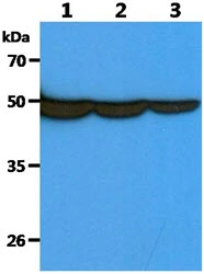
WB analysis of various samples using GTX57621 Fascin 1 antibody. Lane 1 : HeLa whole cell lysate Lane 2 : HepG2 whole cell lysate Lane 3 : THP-1 whole cell lysate Loading : 40 μg Dilution : 1:1000


WB analysis of various samples using GTX57621 Fascin 1 antibody. Lane 1 : HeLa whole cell lysate Lane 2 : HepG2 whole cell lysate Lane 3 : THP-1 whole cell lysate Loading : 40 μg Dilution : 1:1000
Fascin 1 antibody [AT13D2]
GTX57621
ApplicationsFlow Cytometry, ImmunoFluorescence, Western Blot, ImmunoCytoChemistry
Product group Antibodies
ReactivityHuman
TargetFSCN1
Overview
- SupplierGeneTex
- Product NameFascin 1 antibody [AT13D2]
- Delivery Days Customer9
- ApplicationsFlow Cytometry, ImmunoFluorescence, Western Blot, ImmunoCytoChemistry
- CertificationResearch Use Only
- ClonalityMonoclonal
- Clone IDAT13D2
- Concentration1 mg/ml
- ConjugateUnconjugated
- Gene ID6624
- Target nameFSCN1
- Target descriptionfascin actin-bundling protein 1
- Target synonymsFAN1, HSN, SNL, p55, fascin, 55 kDa actin-bundling protein, epididymis secretory sperm binding protein, fascin homolog 1, actin-bundling protein, singed-like (fascin homolog, sea urchin)
- HostMouse
- IsotypeIgG2b
- Protein IDQ16658
- Protein NameFascin
- Scientific DescriptionThis gene encodes a member of the fascin family of actin-binding proteins. Fascin proteins organize F-actin into parallel bundles, and are required for the formation of actin-based cellular protrusions. The encoded protein plays a critical role in cell migration, motility, adhesion and cellular interactions. Expression of this gene is known to be regulated by several microRNAs, and overexpression of this gene may play a role in the metastasis of multiple types of cancer by increasing cell motility. Expression of this gene is also a marker for Reed-Sternberg cells in Hodgkins lymphoma. A pseudogene of this gene is located on the long arm of chromosome 15. [provided by RefSeq, Sep 2011]
- ReactivityHuman
- Storage Instruction-20°C or -80°C,2°C to 8°C
- UNSPSC12352203
Datasheet
Related products
Product group Antibodies

Anti-FSCN1 [SAIC-32C-205]Ab00325-1.1
ApplicationsMass Spectrometry
ReactivityHuman
TargetFSCN1
- SizePrice
Product group Antibodies
Anti-FSCN1 Antibody144-60301
ApplicationsWestern Blot, ImmunoHistoChemistry
ReactivityHuman, Mouse, Rat
TargetFSCN1
- SizePrice
Product group Antibodies
Fascin 1 antibodyGTX117805
ApplicationsImmunoFluorescence, Western Blot, ImmunoCytoChemistry, ImmunoHistoChemistry, ImmunoHistoChemistry Paraffin
ReactivityHuman, Mouse
TargetFSCN1
- SizePrice
![IHC-P analysis of hodgkin's lymphoma tissue using GTX01968 Fascin 1 antibody [IM20].](https://www.genetex.com/upload/website/prouct_img/normal/GTX01968/GTX01968_20200811_IHC-P_37_w_23053121_306.webp)
Product group Antibodies
Fascin 1 antibody [IM20]GTX01968
ApplicationsWestern Blot, ImmunoHistoChemistry, ImmunoHistoChemistry Paraffin
ReactivityHuman
TargetFSCN1
- SizePrice
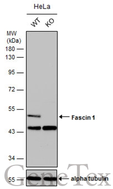
![Wild-type (WT) and Fascin 1 knockout (KO) HeLa cell extracts (30 μg) were separated by 10% SDS-PAGE, and the membrane was blotted with Fascin 1 antibody [N2C2], Internal (GTX100511) diluted at 1:500. The HRP-conjugated anti-rabbit IgG antibody (GTX213110-01) was used to detect the primary antibody, and the signal was developed with Trident ECL plus-Enhanced.](https://www.genetex.com/upload/website/prouct_img/normal/GTX100511/GTX100511_40023_20170406_WB_KO_watermark_w_23060100_778.webp)
Product group Antibodies
References
Fascin 1 antibody [N2C2], InternalGTX100511
ApplicationsWestern Blot
ReactivityHuman
TargetFSCN1
- SizePrice
Product group Antibodies
FSCN1 Polyclonal AntibodyCAC14996
ApplicationsImmunoFluorescence, Western Blot, ELISA, ImmunoHistoChemistry
TargetFSCN1
- SizePrice
Product group Antibodies
References
ApplicationsFlow Cytometry, ImmunoFluorescence, Western Blot, ELISA, ImmunoCytoChemistry, ImmunoHistoChemistry, ImmunoHistoChemistry Frozen, ImmunoHistoChemistry Paraffin
ReactivityCanine, Human, Mouse, Porcine, Rat
TargetFSCN1
- SizePrice
Product group Antibodies
ReactivityHuman
TargetFSCN1
- SizePrice

Product group Antibodies
Anti-FSCN1 AntibodyA97527
ApplicationsWestern Blot, ELISA
ReactivityHuman, Mouse, Rat
- SizePrice