

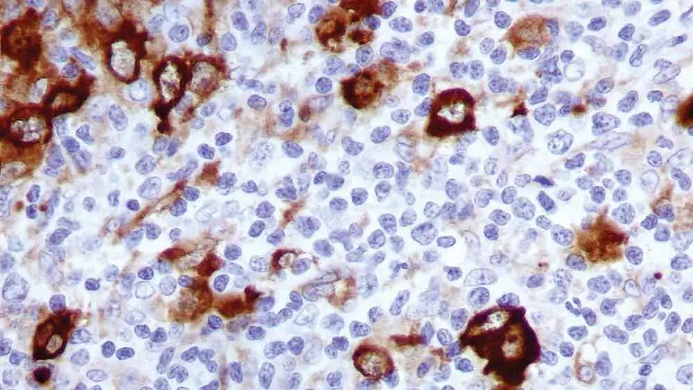
IHC-P analysis of hodgkin's lymphoma tissue using GTX01968 Fascin 1 antibody [IM20].
IHC-P analysis of hodgkin's lymphoma tissue using GTX01968 Fascin 1 antibody [IM20].
Fascin 1 antibody [IM20]
GTX01968
ApplicationsWestern Blot, ImmunoHistoChemistry, ImmunoHistoChemistry Paraffin
Product group Antibodies
ReactivityHuman
TargetFSCN1
Overview
- SupplierGeneTex
- Product NameFascin 1 antibody [IM20]
- Delivery Days Customer9
- Application Supplier NoteWB: 1:100-1:200. IHC-P: 1:200-1:400. *Optimal dilutions/concentrations should be determined by the researcher.Not tested in other applications.
- ApplicationsWestern Blot, ImmunoHistoChemistry, ImmunoHistoChemistry Paraffin
- CertificationResearch Use Only
- ClonalityMonoclonal
- Clone IDIM20
- ConjugateUnconjugated
- Gene ID6624
- Target nameFSCN1
- Target descriptionfascin actin-bundling protein 1
- Target synonymsFAN1, HSN, SNL, p55, fascin, 55 kDa actin-bundling protein, epididymis secretory sperm binding protein, fascin homolog 1, actin-bundling protein, singed-like (fascin homolog, sea urchin)
- HostMouse
- IsotypeIgG1
- Protein IDQ16658
- Protein NameFascin
- Scientific DescriptionThis gene encodes a member of the fascin family of actin-binding proteins. Fascin proteins organize F-actin into parallel bundles, and are required for the formation of actin-based cellular protrusions. The encoded protein plays a critical role in cell migration, motility, adhesion and cellular interactions. Expression of this gene is known to be regulated by several microRNAs, and overexpression of this gene may play a role in the metastasis of multiple types of cancer by increasing cell motility. Expression of this gene is also a marker for Reed-Sternberg cells in Hodgkins lymphoma. A pseudogene of this gene is located on the long arm of chromosome 15. [provided by RefSeq, Sep 2011]
- ReactivityHuman
- Storage Instruction2°C to 8°C
- UNSPSC41116161
Datasheet
Related products

Product group Antibodies
Anti-FSCN1 AntibodyA97527
ApplicationsWestern Blot, ELISA
ReactivityHuman, Mouse, Rat
- SizePrice
Product group Antibodies

Anti-FSCN1 [SAIC-32C-205]Ab00325-1.1
ApplicationsMass Spectrometry
ReactivityHuman
TargetFSCN1
- SizePrice
Product group Antibodies

FSCN1 / Fascin AntibodyLS-C748408
ApplicationsWestern Blot, ImmunoHistoChemistry
ReactivityHuman, Mouse, Rat
TargetFSCN1
- SizePrice
Product group Antibodies
References
ApplicationsFlow Cytometry, ImmunoFluorescence, Western Blot, ELISA, ImmunoCytoChemistry, ImmunoHistoChemistry, ImmunoHistoChemistry Frozen, ImmunoHistoChemistry Paraffin
ReactivityCanine, Human, Mouse, Porcine, Rat
TargetFSCN1
- SizePrice
Product group Antibodies

Goat anti-FSCN1EB08345
ApplicationsWestern Blot, ELISA
ReactivityBovine, Human, Mouse, Rat
TargetFSCN1
- SizePrice
Product group Antibodies
FSCN1 Polyclonal AntibodyCAC14996
ApplicationsImmunoFluorescence, Western Blot, ELISA, ImmunoHistoChemistry
TargetFSCN1
- SizePrice
Product group Antibodies
FSCN1 AntibodyCSB-PA005142
ApplicationsWestern Blot, ELISA
ReactivityHuman, Mouse, Rat
TargetFSCN1
- SizePrice
Product group Antibodies
Fascin 1 antibody [ZM192]GTX01691
ApplicationsImmunoHistoChemistry, ImmunoHistoChemistry Paraffin
ReactivityHuman
TargetFSCN1
- SizePrice
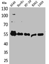
![Wild-type (WT) and Fascin 1 knockout (KO) HeLa cell extracts (30 μg) were separated by 10% SDS-PAGE, and the membrane was blotted with Fascin 1 antibody [N2C2], Internal (GTX100511) diluted at 1:500. The HRP-conjugated anti-rabbit IgG antibody (GTX213110-01) was used to detect the primary antibody, and the signal was developed with Trident ECL plus-Enhanced.](https://www.genetex.com/upload/website/prouct_img/normal/GTX100511/GTX100511_40023_20170406_WB_KO_watermark_w_23060100_778.webp)
Product group Antibodies
Fascin 1 antibody [N2C2], InternalGTX100511
ApplicationsWestern Blot
ReactivityHuman
TargetFSCN1
- SizePrice