

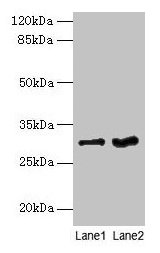
Western blot All lanes: FGFR1OP2 antibody at 6microg/ml Lane 1: Mouse gonadal tissue Lane 2: Human placenta tissue Secondary Goat polyclonal to rabbit IgG at 1/10000 dilution Predicted band size: 30, 25, 21 kDa Observed band size: 30 kDa
Western blot All lanes: FGFR1OP2 antibody at 6microg/ml Lane 1: Mouse gonadal tissue Lane 2: Human placenta tissue Secondary Goat polyclonal to rabbit IgG at 1/10000 dilution Predicted band size: 30, 25, 21 kDa Observed band size: 30 kDa
FGFR1OP2 Antibody
CSB-PA008644LA01HU
ApplicationsWestern Blot, ELISA
Product group Antibodies
ReactivityHuman, Mouse
TargetFGFR1OP2
Overview
- SupplierCusabio
- Product NameFGFR1OP2 Antibody
- Delivery Days Customer20
- ApplicationsWestern Blot, ELISA
- CertificationResearch Use Only
- ClonalityPolyclonal
- ConjugateUnconjugated
- Gene ID26127
- Target nameFGFR1OP2
- Target descriptionFGFR1 oncogene partner 2
- Target synonymsHSPC123-like, WIT3.0, FGFR1 oncogene partner 2, fibroblast growth factor receptor 1 oncogene partner 2, wound inducible transcript 3.0
- HostRabbit
- IsotypeIgG
- Protein IDQ9NVK5
- Protein NameFGFR1 oncogene partner 2
- Scientific DescriptionMay be involved in wound healing pathway.
- ReactivityHuman, Mouse
- Storage Instruction-20°C or -80°C
- UNSPSC41116161
Related products
Product group Antibodies
Anti-FGFR1OP2 AntibodyA44997
ApplicationsImmunoHistoChemistry
ReactivityHuman
- SizePrice
Product group Antibodies
Anti-FGFR1OP2 Antibody102-20222
ApplicationsFlow Cytometry, Western Blot
TargetFGFR1OP2
- SizePrice

Product group Antibodies
Anti-FGFR1OP2 AntibodyM10171
ApplicationsFlow Cytometry
ReactivityHuman, Mouse
TargetFGFR1OP2
- SizePrice
Product group Antibodies
FGFR1OP2 Polyclonal AntibodyBS-8346R
ApplicationsImmunoFluorescence, Western Blot, ELISA, ImmunoCytoChemistry, ImmunoHistoChemistry, ImmunoHistoChemistry Frozen, ImmunoHistoChemistry Paraffin
ReactivityCanine, Equine, Human, Mouse, Rabbit, Rat
TargetFGFR1OP2
- SizePrice
Product group Antibodies
Fgfr1Op2 Polyclonal AntibodyCAC08437
ApplicationsWestern Blot, ELISA
ReactivityMouse
TargetFGFR1OP2
- SizePrice
Product group Antibodies

FGFR1OP2 AntibodyLS-C401678
ApplicationsELISA, ImmunoHistoChemistry
ReactivityHuman, Mouse, Rat
TargetFGFR1OP2
- SizePrice
Product group Antibodies
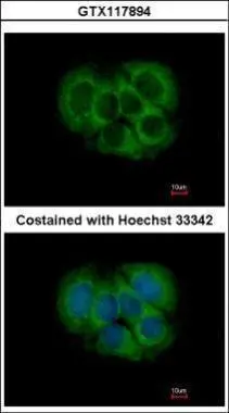
FGFR1OP2 antibodyGTX117894
ApplicationsImmunoFluorescence, Western Blot, ImmunoCytoChemistry, ImmunoHistoChemistry, ImmunoHistoChemistry Paraffin
ReactivityHuman, Mouse
TargetFGFR1OP2
- SizePrice

Product group Antibodies
Anti-FGFR1OP2 AntibodyHPA038308
ApplicationsWestern Blot, ImmunoHistoChemistry
ReactivityHuman
TargetFGFR1OP2
- SizePrice