

FGFR1OP2 Polyclonal Antibody
BS-8346R
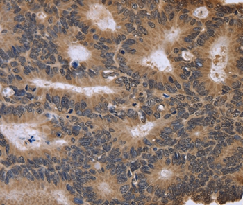
ApplicationsImmunoFluorescence, Western Blot, ELISA, ImmunoCytoChemistry, ImmunoHistoChemistry, ImmunoHistoChemistry Frozen, ImmunoHistoChemistry Paraffin
Product group Antibodies
ReactivityCanine, Equine, Human, Mouse, Rabbit, Rat
TargetFGFR1OP2
Overview
- SupplierBioss
- Product NameFGFR1OP2 Polyclonal Antibody
- Delivery Days Customer7
- ApplicationsImmunoFluorescence, Western Blot, ELISA, ImmunoCytoChemistry, ImmunoHistoChemistry, ImmunoHistoChemistry Frozen, ImmunoHistoChemistry Paraffin
- Applications SupplierWB(1:300-5000), ELISA(1:500-1000), IHC-P(1:200-400), IHC-F(1:100-500), IF(IHC-P)(1:50-200), IF(IHC-F)(1:50-200), IF(ICC)(1:50-200)
- CertificationResearch Use Only
- ClonalityPolyclonal
- Concentration1 ug/ul
- ConjugateUnconjugated
- Gene ID26127
- Target nameFGFR1OP2
- Target descriptionFGFR1 oncogene partner 2
- Target synonymsHSPC123-like, WIT3.0, FGFR1 oncogene partner 2, fibroblast growth factor receptor 1 oncogene partner 2, wound inducible transcript 3.0
- HostRabbit
- IsotypeIgG
- ReactivityCanine, Equine, Human, Mouse, Rabbit, Rat
- Storage Instruction-20°C
- UNSPSC41116161
Related products
Product group Antibodies
Anti-FGFR1OP2 AntibodyA44997
ApplicationsImmunoHistoChemistry
ReactivityHuman
- SizePrice
Product group Antibodies
Anti-FGFR1OP2 Antibody102-20222
ApplicationsFlow Cytometry, Western Blot
TargetFGFR1OP2
- SizePrice

Product group Antibodies
Anti-FGFR1OP2 AntibodyM10171
ApplicationsFlow Cytometry
ReactivityHuman, Mouse
TargetFGFR1OP2
- SizePrice
Product group Antibodies
FGFR1OP2 AntibodyCSB-PA008644LA01HU
ApplicationsWestern Blot, ELISA
ReactivityHuman, Mouse
TargetFGFR1OP2
- SizePrice
Product group Antibodies
Fgfr1Op2 Polyclonal AntibodyCAC08437
ApplicationsWestern Blot, ELISA
ReactivityMouse
TargetFGFR1OP2
- SizePrice
Product group Antibodies

FGFR1OP2 AntibodyLS-C401678
ApplicationsELISA, ImmunoHistoChemistry
ReactivityHuman, Mouse, Rat
TargetFGFR1OP2
- SizePrice
Product group Antibodies
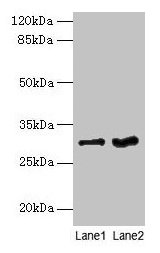
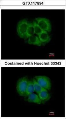
FGFR1OP2 antibodyGTX117894
ApplicationsImmunoFluorescence, Western Blot, ImmunoCytoChemistry, ImmunoHistoChemistry, ImmunoHistoChemistry Paraffin
ReactivityHuman, Mouse
TargetFGFR1OP2
- SizePrice

Product group Antibodies
Anti-FGFR1OP2 AntibodyHPA038308
ApplicationsWestern Blot, ImmunoHistoChemistry
ReactivityHuman
TargetFGFR1OP2
- SizePrice