

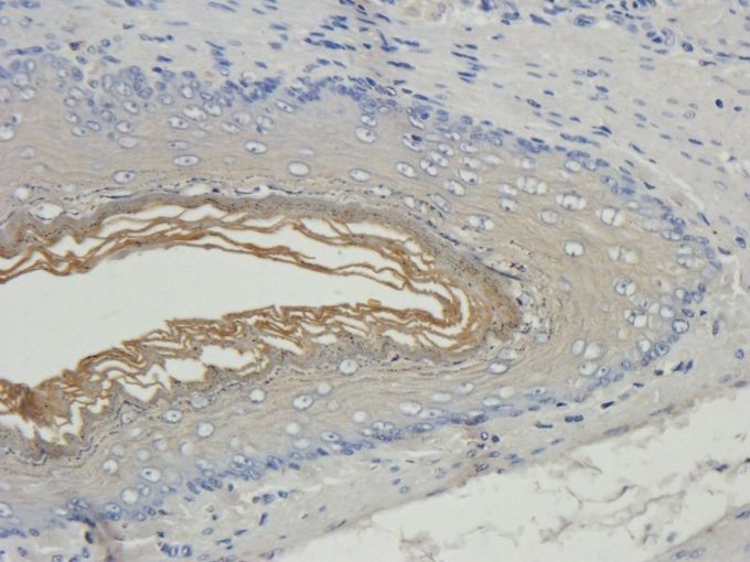
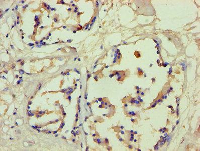
GTX37695 IHC-P Image


GTX37695 IHC-P Image
Filaggrin antibody
GTX37695
ApplicationsImmunoHistoChemistry, ImmunoHistoChemistry Paraffin
Product group Antibodies
ReactivityHuman, Mouse, Rat
TargetFLG
Overview
- SupplierGeneTex
- Product NameFilaggrin antibody
- Delivery Days Customer9
- Application Supplier NoteIHC-P: 1:100-500 (based on 0.5 mg/ml). *Optimal dilutions/concentrations should be determined by the researcher.Not tested in other applications.
- ApplicationsImmunoHistoChemistry, ImmunoHistoChemistry Paraffin
- CertificationResearch Use Only
- ClonalityPolyclonal
- Concentration0.5 mg/ml
- ConjugateUnconjugated
- Gene ID2312
- Target nameFLG
- Target descriptionfilaggrin
- Target synonymsATOD2, FLG-1, FLG1, filaggrin, epidermal filaggrin
- HostRabbit
- IsotypeIgG
- Protein IDP20930
- Protein NameFilaggrin
- Scientific DescriptionThe protein encoded by this gene is an intermediate filament-associated protein that aggregates keratin intermediate filaments in mammalian epidermis. It is initially synthesized as a polyprotein precursor, profilaggrin (consisting of multiple filaggrin units of 324 aa each), which is localized in keratohyalin granules, and is subsequently proteolytically processed into individual functional filaggrin molecules. Mutations in this gene are associated with ichthyosis vulgaris.[provided by RefSeq, Dec 2009]
- ReactivityHuman, Mouse, Rat
- Storage Instruction-20°C or -80°C,2°C to 8°C
- UNSPSC41116161
References
- Effects of Oral Administration of Lactiplantibacillus Plantarum APsulloc 331261 (GTB1(TM)) Isolated from Green Tea on Atopic Dermatitis (AD)-like Skin Lesion Mouse Models.Read this paper
- Effects of chloroform fraction of Fritillariae Thunbergii Bulbus on atopic symptoms in a DNCB-induced atopic dermatitis-like skin lesion model and in vitro models. Kim EY et al., 2021 Dec 5, J EthnopharmacolRead this paper
- Chondroitin 6-sulfate represses keratinocyte proliferation in mouse skin, which is associated with psoriasis. Kitazawa K et al., 2021 Jan 25, Commun BiolRead this paper
- Glycofullerenes Inhibit Particulate Matter Induced Inflammation and Loss of Barrier Proteins in HaCaT Human Keratinocytes. Lee CW et al., 2020 Mar 28, BiomoleculesRead this paper
- Fermented Morinda citrifolia (Noni) Alleviates DNCB-Induced Atopic Dermatitis in NC/Nga Mice through Modulating Immune Balance and Skin Barrier Function. Kim SH et al., 2020 Jan 18, NutrientsRead this paper
- Evidence for the critical role of the PI3K signaling pathway in particulate matter-induced dysregulation of the inflammatory mediators COX-2/PGE2 and the associated epithelial barrier protein Filaggrin in the bronchial epithelium. Song C et al., 2020 Aug, Cell Biol ToxicolRead this paper
- Water-Soluble Fullerenol C60(OH)36 toward Effective Anti-Air Pollution Induced by Urban Particulate Matter in HaCaT Cell. Lee CW et al., 2019 Aug 30, Int J Mol SciRead this paper
- Borage oil restores acidic skin pH by up-regulating the activity or expression of filaggrin and enzymes involved in epidermal lactate, free fatty acid, and acidic free amino acid metabolism in essential fatty acid-deficient Guinea pigs. Kim KP et al., 2018 Oct, Nutr ResRead this paper
Datasheet
Related products
Product group Antibodies
FLG AntibodyCSB-PA008712HA01HU
ApplicationsELISA, ImmunoHistoChemistry
ReactivityHuman
TargetFLG
- SizePrice
Product group Antibodies
Anti-Filaggrin AntibodyA28578
ApplicationsWestern Blot
ReactivityHuman, Mouse, Rat
- SizePrice
Product group Antibodies

FLG / Filaggrin AntibodyLS-C783376
ApplicationsWestern Blot
ReactivityHuman
TargetFLG
- SizePrice

Product group Antibodies
Anti-FLG AntibodyHPA030188
ApplicationsImmunoCytoChemistry, ImmunoHistoChemistry
ReactivityHuman
TargetFLG
- SizePrice
![IHC-P analysis of human skin tissue using GTX34731 Filaggrin antibody [FLG/1561].](https://www.genetex.com/upload/website/prouct_img/normal/GTX34731/GTX34731_20200115_IHC-P_1125_w_23060801_149.webp)
Product group Antibodies
Filaggrin antibody [FLG/1561]GTX34731
ApplicationsImmunoHistoChemistry, ImmunoHistoChemistry Paraffin, Other Application
ReactivityHuman
TargetFLG
- SizePrice
![IHC-P analysis of human skin tissue using GTX34732 Filaggrin antibody [SPM181].](https://www.genetex.com/upload/website/prouct_img/normal/GTX34732/GTX34732_20200115_IHC-P_1124_w_23060801_185.webp)
Product group Antibodies
References
Filaggrin antibody [SPM181]GTX34732
ApplicationsImmunoHistoChemistry, ImmunoHistoChemistry Paraffin
ReactivityHuman
TargetFLG
- SizePrice
![IHC-P analysis of human skin tissue using GTX34733 Filaggrin antibody [FLG/1562].](https://www.genetex.com/upload/website/prouct_img/normal/GTX34733/GTX34733_20200115_IHC-P_1126_w_23060801_695.webp)
Product group Antibodies
Filaggrin antibody [FLG/1562]GTX34733
ApplicationsImmunoHistoChemistry, ImmunoHistoChemistry Paraffin
ReactivityHuman
TargetFLG
- SizePrice

Product group Antibodies
Anti-Filaggrin/FLG Antibody Picoband(r)PB9870-CARRIER-FREE
ApplicationsWestern Blot, ImmunoHistoChemistry
ReactivityHuman
TargetFLG
- SizePrice
![IHC-P analysis of human skin tissue section using GTX02638 Filaggrin antibody [FLG/1957R].](https://www.genetex.com/upload/website/prouct_img/normal/GTX02638/GTX02638_20210319_IHC-P_w_23053122_509.webp)
Product group Antibodies
Filaggrin antibody [FLG/1957R]GTX02638
ApplicationsImmunoHistoChemistry, ImmunoHistoChemistry Paraffin
ReactivityHuman
TargetFLG
- SizePrice