

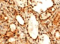
IHC-P analysis of human thyroid Gland using GTX25080 FOXE1 antibody, C-term. Antigen retrieval : Tris/EDTA buffer pH 9 Dilution : 2μg/ml

IHC-P analysis of human thyroid Gland using GTX25080 FOXE1 antibody, C-term. Antigen retrieval : Tris/EDTA buffer pH 9 Dilution : 2μg/ml
FOXE1 antibody, C-term
GTX25080
ApplicationsImmunoHistoChemistry, ImmunoHistoChemistry Paraffin
Product group Antibodies
ReactivityHuman
TargetFOXE1
Overview
- SupplierGeneTex
- Product NameFOXE1 antibody, C-term
- Delivery Days Customer9
- Application Supplier NoteIHC-P: 5microg/ml. *Optimal dilutions/concentrations should be determined by the researcher.Not tested in other applications.
- ApplicationsImmunoHistoChemistry, ImmunoHistoChemistry Paraffin
- CertificationResearch Use Only
- ClonalityPolyclonal
- Concentration0.50 mg/ml
- ConjugateUnconjugated
- Gene ID2304
- Target nameFOXE1
- Target descriptionforkhead box E1
- Target synonymsBAMLAZ, FKHL15, FOXE2, HFKH4, HFKL5, NMTC4, TITF2, TTF-2, TTF2, forkhead box protein E1, HNF-3/fork head-like protein 5, forkhead box protein E2, forkhead, drosophila, homolog-like 15, forkhead-related protein FKHL15, thyroid transcription factor 2
- HostGoat
- IsotypeIgG
- Protein IDO00358
- Protein NameForkhead box protein E1
- Scientific DescriptionThis intronless gene encodes a protein that belongs to the forkhead family of transcription factors. Members of this family contain a conserved 100-amino acid DNA-binding forkhead domain. The encoded protein functions as a thyroid transcription factor that plays a role in thyroid morphogenesis. Mutations in this gene are associated with the Bamforth-Lazarus syndrome, and with susceptibility to nonmedullary thyroid cancer-4. [provided by RefSeq, Nov 2016]
- ReactivityHuman
- Storage Instruction-20°C or -80°C,2°C to 8°C
- UNSPSC41116161
Datasheet
Related products
Product group Antibodies
FOXE1 AntibodyCSB-PA008314
ApplicationsImmunoFluorescence, ELISA
ReactivityHuman, Mouse, Rat
TargetFOXE1
- SizePrice
Product group Antibodies
Anti-TTF2 AntibodyA97875
ApplicationsWestern Blot, ELISA
ReactivityHuman, Mouse, Rat
- SizePrice

Product group Antibodies
Anti-FOXE1 Picoband(r) AntibodyA02831-5-CARRIER-FREE
ApplicationsFlow Cytometry, Western Blot, ELISA
ReactivityHuman
TargetFOXE1
- SizePrice
Product group Antibodies

FOXE2 / FOXE1 AntibodyLS-C747721
ApplicationsWestern Blot
ReactivityHuman
TargetFOXE1
- SizePrice
Product group Antibodies

Goat anti-FOXE1 / TTF2EB06011
ApplicationsELISA, ImmunoHistoChemistry
ReactivityHuman
TargetFOXE1
- SizePrice
Product group Antibodies
Foxe1 Polyclonal AntibodyCAC08156
ApplicationsImmunoFluorescence, Western Blot, ELISA
ReactivityMouse
TargetFOXE1
- SizePrice
Product group Antibodies
FOXE1 antibodyGTX100275
ApplicationsImmunoPrecipitation, Western Blot
ReactivityHuman
TargetFOXE1
- SizePrice
Product group Antibodies
References
FOXE1 Polyclonal AntibodyBS-0446R
ApplicationsImmunoFluorescence, Western Blot, ELISA, ImmunoCytoChemistry, ImmunoHistoChemistry, ImmunoHistoChemistry Frozen, ImmunoHistoChemistry Paraffin
ReactivityBovine, Canine, Chicken, Human, Mouse, Porcine, Rat
TargetFOXE1
- SizePrice

Product group Antibodies
FOXE1 antibodyGTX17612
ApplicationsWestern Blot
ReactivityHuman
TargetFOXE1
- SizePrice