

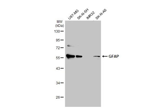
Various whole cell extracts (30 μg) were separated by 10% SDS-PAGE, and the membrane was blotted with GFAP antibody (GTX108711) diluted at 1:2500. The HRP-conjugated anti-rabbit IgG antibody (GTX213110-01) was used to detect the primary antibody.
![GFAP antibody detects GFAP protein at glia cells by immunofluorescent analysis. Sample: DIV10 rat E18 primary hippocampal neuron and glia cells were fixed in 4% paraformaldehyde at RT for 15 min. Green: GFAP stained by GFAP antibody (GTX108711) diluted at 1:250. Red: Tau, a cytoskeleton marker, stained by Tau antibody [GT287] (GTX634809) diluted at 1:500. Blue: Fluoroshield with DAPI (GTX30920).](https://www.genetex.com/upload/website/prouct_img/normal/GTX108711/GTX108711_44615_20220916_ICC_IF_R_22110201_470.webp "GFAP antibody detects GFAP protein at glia cells by immunofluorescent analysis. Sample: DIV10 rat E18 primary hippocampal neuron and glia cells were fixed in 4% paraformaldehyde at RT for 15 min. Green: GFAP stained by GFAP antibody (GTX108711) diluted at 1:250. Red: Tau, a cytoskeleton marker, stained by Tau antibody [GT287] (GTX634809) diluted at 1:500. Blue: Fluoroshield with DAPI (GTX30920).")
 were separated by 10% SDS-PAGE, and the membrane was blotted with GFAP antibody (GTX108711) diluted at 1:25000. The HRP-conjugated anti-rabbit IgG antibody (GTX213110-01) was used to detect the primary antibody.")
 and transfected (+) 293T whole cell extracts (30 μg) were separated by 10% SDS-PAGE, and the membrane was blotted with GFAP antibody (GTX108711) diluted at 1:20000. The HRP-conjugated anti-rabbit IgG antibody (GTX213110-01) was used to detect the primary antibody.")
 diluted at 1:500. Antigen Retrieval: Citrate buffer, pH 6.0, 15 min")
 diluted at 1:500. Antigen Retrieval: Citrate buffer, pH 6.0, 15 min")
![GFAP antibody detects GFAP protein expression by immunohistochemical analysis. Sample: Frozen-sectioned adult mouse hippocampus. Green: GFAP protein stained by GFAP antibody (GTX108711) diluted at 1:250. Red: NeuN, stained by NeuN antibody [2Q158] (GTX30773) diluted at 1:500.](https://www.genetex.com/upload/website/prouct_img/normal/GTX108711/GTX108711_39981_42886_IHC-Fr_M_w_23060120_269.webp "GFAP antibody detects GFAP protein expression by immunohistochemical analysis. Sample: Frozen-sectioned adult mouse hippocampus. Green: GFAP protein stained by GFAP antibody (GTX108711) diluted at 1:250. Red: NeuN, stained by NeuN antibody [2Q158] (GTX30773) diluted at 1:500.")
 diluted at 1:200. The signal was developed using goat anti-rabbit IgG antibody (Dylight488) (GTX213110-04). Blue: Nuclear staining with Hoechst 33342.
Antigen Retrieval: Citrate buffer, pH 6.0, 15 min")
![GFAP antibody detects GFAP protein at glia cells by immunofluorescent analysis. Sample: DIV10 rat E18 primary cortical neuron and glia cells were fixed in 4% paraformaldehyde at RT for 15 min. Green: GFAP stained by GFAP antibody (GTX108711) diluted at 1:500. Red: Tau, stained by Tau antibody [GT287] (GTX634809) diluted at 1:500. Blue: Fluoroshield with DAPI (GTX30920).](https://www.genetex.com/upload/website/prouct_img/normal/GTX108711/GTX108711_43596_20191125_ICC_IF_R_w_23060120_219.webp "GFAP antibody detects GFAP protein at glia cells by immunofluorescent analysis. Sample: DIV10 rat E18 primary cortical neuron and glia cells were fixed in 4% paraformaldehyde at RT for 15 min. Green: GFAP stained by GFAP antibody (GTX108711) diluted at 1:500. Red: Tau, stained by Tau antibody [GT287] (GTX634809) diluted at 1:500. Blue: Fluoroshield with DAPI (GTX30920).")
 diluted at 1:300. Blue: Hoechst 33342 staining.")
Various whole cell extracts (30 μg) were separated by 10% SDS-PAGE, and the membrane was blotted with GFAP antibody (GTX108711) diluted at 1:2500. The HRP-conjugated anti-rabbit IgG antibody (GTX213110-01) was used to detect the primary antibody.
GFAP antibody
GTX108711
ApplicationsImmunoFluorescence, Western Blot, ImmunoCytoChemistry, ImmunoHistoChemistry, ImmunoHistoChemistry Frozen, ImmunoHistoChemistry Paraffin
Product group Antibodies
ReactivityHuman, Mouse, Rat
TargetGFAP
Overview
- SupplierGeneTex
- Product NameGFAP antibody
- Delivery Days Customer9
- Application Supplier NoteWB: 1:5000-1:50000. ICC/IF: 1:100-1:1000. IHC-P: 1:100-1:1000. IHC-Fr: 1:100-1:1000. *Optimal dilutions/concentrations should be determined by the researcher.Not tested in other applications.
- ApplicationsImmunoFluorescence, Western Blot, ImmunoCytoChemistry, ImmunoHistoChemistry, ImmunoHistoChemistry Frozen, ImmunoHistoChemistry Paraffin
- CertificationResearch Use Only
- ClonalityPolyclonal
- Concentration0.47 mg/ml
- ConjugateUnconjugated
- Gene ID2670
- Target nameGFAP
- Target descriptionglial fibrillary acidic protein
- Target synonymsALXDRD, glial fibrillary acidic protein
- HostRabbit
- IsotypeIgG
- Protein IDP14136
- Protein NameGlial fibrillary acidic protein
- Scientific DescriptionThis gene encodes one of the major intermediate filament proteins of mature astrocytes. It is used as a marker to distinguish astrocytes from other glial cells during development. Mutations in this gene cause Alexander disease, a rare disorder of astrocytes in the central nervous system. Alternative splicing results in multiple transcript variants encoding distinct isoforms. [provided by RefSeq]
- ReactivityHuman, Mouse, Rat
- Storage Instruction-20°C or -80°C,2°C to 8°C
- UNSPSC12352203
References
- Sun MH, Ho TC, Yeh SI, et al. Short peptides derived from pigment epithelium-derived factor attenuate retinal ischemia reperfusion injury through inhibition of apoptosis and inflammatory response in rats. Exp Eye Res. 2024,238:109743. doi: 10.1016/j.exer.2023.109743Read this paper
- Wei H, Zhen L, Wang S, et al. Glyceryl triacetate promotes blood-brain barrier recovery after ischemic stroke through lipogenesis-mediated IL-33 in mice. J Neuroinflammation. 2023,20(1):264. doi: 10.1186/s12974-023-02942-3Read this paper
- Ayon-Olivas M, Wolf D, Andreska T, et al. Dopaminergic Input Regulates the Sensitivity of Indirect Pathway Striatal Spiny Neurons to Brain-Derived Neurotrophic Factor. Biology (Basel). 2023,12(10). doi: 10.3390/biology12101360Read this paper
- Gimeno-Ferrer F, Eitner A, Bauer R, et al. Cortical spreading depolarization is a potential target for rat brain excitability modulation by Galanin. Exp Neurol. 2023,370:114569. doi: 10.1016/j.expneurol.2023.114569Read this paper
- Chiu YS, Wu KJ, Yu SJ, et al. Peptide immunization against the C-terminal of alpha-synuclein reduces locomotor activity in mice overexpressing alpha-synuclein. PLoS One. 2023,18(9):e0291927. doi: 10.1371/journal.pone.0291927Read this paper
- Kun W, Jie Z, Shuai C, et al. Electroacupuncture ameliorates cardiac dysfunction in myocardial ischemia model rats: a potential role of the hypothalamic-pituitary-adrenal axis. J Tradit Chin Med. 2023,43(5):944-954. doi: 10.19852/j.cnki.jtcm.20230727.001Read this paper
- Makibatake R, Oda S, Yagi Y, et al. Amyloid-β slows cilia movement along the ventricle, impairs fluid flow, and exacerbates its neurotoxicity in explant culture. Sci Rep. 2023,13(1):13586. doi: 10.1038/s41598-023-40742-0Read this paper
- Montillot C, Skutunova E, Ayushma, et al. Characterization of Vps13b-mutant mice reveals neuroanatomical and behavioral phenotypes with females less affected. Neurobiol Dis. 2023,185:106259. doi: 10.1016/j.nbd.2023.106259Read this paper
- Kobayashi M, Moro N, Yoshino A, et al. Inhibition of P2X4 and P2X7 receptors improves histological and behavioral outcomes after experimental traumatic brain injury in rats. Exp Ther Med. 2023,26(2):378. doi: 10.3892/etm.2023.12077Read this paper
- Liang MZ, Lu TH, Chen L. Timely expression of PGAM5 and its cleavage control mitochondrial homeostasis during neurite re-growth after traumatic brain injury. Cell Biosci. 2023,13(1):96. doi: 10.1186/s13578-023-01052-0Read this paper
Datasheet
Related products
Product group Antibodies

Anti-GFAP R416WT [N206B/9]Ab02152-1.1
ApplicationsWestern Blot, ImmunoCytoChemistry, ImmunoHistoChemistry
ReactivityHuman, Mouse, Rat
TargetGFAP
- SizePrice
Product group Antibodies
Anti-GFAP Antibody130-10037
ApplicationsELISA
ReactivityHuman
TargetGFAP
- SizePrice

Product group Antibodies
Anti-GFAP AntibodyAMAB91033
ApplicationsWestern Blot, ImmunoHistoChemistry
ReactivityHuman, Mouse, Rat
TargetGFAP
- SizePrice
Product group Antibodies

ApplicationsImmunoFluorescence, Western Blot, ELISA, ImmunoCytoChemistry, ImmunoHistoChemistry
- SizePrice
Product group Antibodies
References
GFAP antibodyGTX100850
ApplicationsImmunoFluorescence, Western Blot, ImmunoCytoChemistry, ImmunoHistoChemistry, ImmunoHistoChemistry Frozen, ImmunoHistoChemistry Paraffin
ReactivityHuman, Mouse, Rat
TargetGFAP
- SizePrice
![GFAP antibody [HL1307] detects GFAP protein at cytoplasm by immunohistochemical analysis. Sample: Paraffin-embedded mouse brain. GFAP stained by GFAP antibody [HL1307] (GTX636725) diluted at 1:100. Antigen Retrieval: Citrate buffer, pH 6.0, 15 min](https://www.genetex.com/upload/website/prouct_img/normal/GTX636725/GTX636725_44627_20220408_IHC-P_M_22071401_482.webp)
Product group Antibodies
GFAP antibody [HL1307]GTX636725
ApplicationsWestern Blot, ImmunoHistoChemistry, ImmunoHistoChemistry Frozen, ImmunoHistoChemistry Paraffin
ReactivityHuman, Mouse, Rat
TargetGFAP
- SizePrice
![GFAP antibody [HL1308] detects GFAP protein at cytoplasm by immunohistochemical analysis. Sample: Paraffin-embedded rat brain. GFAP stained by GFAP antibody [HL1308] (GTX636726) diluted at 1:100. Antigen Retrieval: Citrate buffer, pH 6.0, 15 min](https://www.genetex.com/upload/website/prouct_img/normal/GTX636726/GTX636726_44627_20220408_IHC-P_R_22071401_888.webp)
Product group Antibodies
GFAP antibody [HL1308]GTX636726
ApplicationsImmunoFluorescence, ImmunoCytoChemistry, ImmunoHistoChemistry, ImmunoHistoChemistry Frozen, ImmunoHistoChemistry Paraffin
ReactivityHuman, Mouse, Rat
TargetGFAP
- SizePrice
Product group Antibodies
References
GFAP antibodyGTX85454
ApplicationsImmunoFluorescence, Western Blot, ImmunoCytoChemistry, ImmunoHistoChemistry, ImmunoHistoChemistry Frozen, ImmunoHistoChemistry Paraffin
ReactivityHuman, Mouse, Rat
TargetGFAP
- SizePrice
Product group Antibodies
Gfap Polyclonal AntibodyCAC08463
ApplicationsImmunoFluorescence, Western Blot, ELISA, ImmunoHistoChemistry
TargetGFAP
- SizePrice