

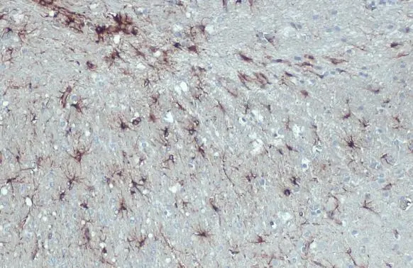
GFAP antibody [HL1308] detects GFAP protein at cytoplasm by immunohistochemical analysis. Sample: Paraffin-embedded rat brain. GFAP stained by GFAP antibody [HL1308] (GTX636726) diluted at 1:100. Antigen Retrieval: Citrate buffer, pH 6.0, 15 min
![GFAP antibody [HL1308] detects GFAP protein at glia cells by immunofluorescent analysis. Sample: DIV10 rat E18 primary hippocampal neuron and glia cells were fixed in 4% paraformaldehyde at RT for 15 min. Green: GFAP stained by GFAP antibody [HL1308] (GTX636726) diluted at 1:250. Red: Tau, a cytoskeleton marker, stained by Tau antibody [GT287] (GTX634809) diluted at 1:500. Blue: Fluoroshield with DAPI (GTX30920).](https://www.genetex.com/upload/website/prouct_img/normal/GTX636726/GTX636726_44627_20220916_ICC_IF_R_22102723_936.webp "GFAP antibody [HL1308] detects GFAP protein at glia cells by immunofluorescent analysis. Sample: DIV10 rat E18 primary hippocampal neuron and glia cells were fixed in 4% paraformaldehyde at RT for 15 min. Green: GFAP stained by GFAP antibody [HL1308] (GTX636726) diluted at 1:250. Red: Tau, a cytoskeleton marker, stained by Tau antibody [GT287] (GTX634809) diluted at 1:500. Blue: Fluoroshield with DAPI (GTX30920).")
![GFAP antibody [HL1308] detects GFAP protein by immunohistochemical analysis. Sample: Frozen-sectioned mouse cerebellum. Green: GFAP stained by GFAP antibody [HL1308] (GTX636726) diluted at 1:100.](https://www.genetex.com/upload/website/prouct_img/normal/GTX636726/GTX636726_44627_20221202_IHC-Fr_M_22121123_898.webp "GFAP antibody [HL1308] detects GFAP protein by immunohistochemical analysis. Sample: Frozen-sectioned mouse cerebellum. Green: GFAP stained by GFAP antibody [HL1308] (GTX636726) diluted at 1:100.")
![GFAP antibody [HL1308] detects GFAP protein at glia cells by immunofluorescent analysis. Sample: DIV9 rat E18 primary cortical neuron and glia cells were fixed in 4% paraformaldehyde at RT for 15 min. Green: GFAP stained by GFAP antibody [HL1308] (GTX636726) diluted at 1:250. Red: Tau, an axon marker, stained by Tau antibody [GT287] (GTX634809) diluted at 1:500. Blue: Fluoroshield with DAPI (GTX30920).](https://www.genetex.com/upload/website/prouct_img/normal/GTX636726/GTX636726_44627_20221209_ICC_IF_R_22122018_760.webp "GFAP antibody [HL1308] detects GFAP protein at glia cells by immunofluorescent analysis. Sample: DIV9 rat E18 primary cortical neuron and glia cells were fixed in 4% paraformaldehyde at RT for 15 min. Green: GFAP stained by GFAP antibody [HL1308] (GTX636726) diluted at 1:250. Red: Tau, an axon marker, stained by Tau antibody [GT287] (GTX634809) diluted at 1:500. Blue: Fluoroshield with DAPI (GTX30920).")
![GFAP antibody [HL1308] detects GFAP protein at cytoplasm by immunohistochemical analysis. Sample: Frozen-sectioned mouse spinal cord. Green: GFAP stained by GFAP antibody [HL1308] (GTX636726) diluted at 1:100. Blue: Fluoroshield with DAPI (GTX30920).](https://www.genetex.com/upload/website/prouct_img/normal/GTX636726/GTX636726_44627_20230414_IHC-Fr_M_23041719_696.webp "GFAP antibody [HL1308] detects GFAP protein at cytoplasm by immunohistochemical analysis. Sample: Frozen-sectioned mouse spinal cord. Green: GFAP stained by GFAP antibody [HL1308] (GTX636726) diluted at 1:100. Blue: Fluoroshield with DAPI (GTX30920).")
![GFAP antibody [HL1308] detects GFAP protein at cytoplasm by immunohistochemical analysis. Sample: Paraffin-embedded rat brain. GFAP stained by GFAP antibody [HL1308] (GTX636726) diluted at 1:100. Antigen Retrieval: Citrate buffer, pH 6.0, 15 min](https://www.genetex.com/upload/website/prouct_img/normal/GTX636726/GTX636726_T-44564_20220128_IHC-P_R_w_23061202_988.webp "GFAP antibody [HL1308] detects GFAP protein at cytoplasm by immunohistochemical analysis. Sample: Paraffin-embedded rat brain. GFAP stained by GFAP antibody [HL1308] (GTX636726) diluted at 1:100. Antigen Retrieval: Citrate buffer, pH 6.0, 15 min")
![GFAP antibody [HL1308] detects GFAP protein at glia cells by immunohistochemical analysis. Sample: Frozen-sectioned mouse brain. Green: GFAP stained by GFAP antibody [HL1308] (GTX636726) diluted at 1:200. Blue: Fluoroshield with DAPI (GTX30920).](https://www.genetex.com/upload/website/prouct_img/normal/GTX636726/GTX636726_44627_20220429_IHC-Fr_M_w_23061202_414.webp "GFAP antibody [HL1308] detects GFAP protein at glia cells by immunohistochemical analysis. Sample: Frozen-sectioned mouse brain. Green: GFAP stained by GFAP antibody [HL1308] (GTX636726) diluted at 1:200. Blue: Fluoroshield with DAPI (GTX30920).")
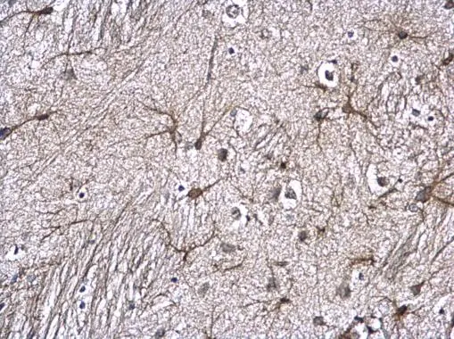
![GFAP antibody [HL1308] detects GFAP protein by immunohistochemical analysis. Sample: Paraffin-embedded mouse hippocampus. GFAP stained by GFAP antibody [HL1308] (GTX636726) diluted at 1:100. Antigen Retrieval: Citrate buffer, pH 6.0, 15 min](https://www.genetex.com/upload/website/prouct_img/normal/GTX636726/GTX636726_45719_20250429_IHC-P_M_25051420_899.webp "GFAP antibody [HL1308] detects GFAP protein by immunohistochemical analysis. Sample: Paraffin-embedded mouse hippocampus. GFAP stained by GFAP antibody [HL1308] (GTX636726) diluted at 1:100. Antigen Retrieval: Citrate buffer, pH 6.0, 15 min")
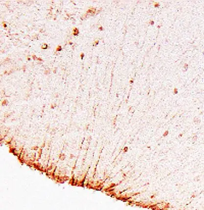
GFAP antibody [HL1308] detects GFAP protein at cytoplasm by immunohistochemical analysis. Sample: Paraffin-embedded rat brain. GFAP stained by GFAP antibody [HL1308] (GTX636726) diluted at 1:100. Antigen Retrieval: Citrate buffer, pH 6.0, 15 min
GFAP antibody [HL1308]
GTX636726
ApplicationsImmunoFluorescence, ImmunoCytoChemistry, ImmunoHistoChemistry, ImmunoHistoChemistry Frozen, ImmunoHistoChemistry Paraffin
Product group Antibodies
ReactivityHuman, Mouse, Rat
TargetGFAP
Overview
- SupplierGeneTex
- Product NameGFAP antibody [HL1308]
- Delivery Days Customer9
- Application Supplier NoteIHC-P: 1:100-1:1000. *Optimal dilutions/concentrations should be determined by the researcher.Not tested in other applications.
- ApplicationsImmunoFluorescence, ImmunoCytoChemistry, ImmunoHistoChemistry, ImmunoHistoChemistry Frozen, ImmunoHistoChemistry Paraffin
- CertificationResearch Use Only
- ClonalityMonoclonal
- Clone IDHL1308
- Concentration1 mg/ml
- ConjugateUnconjugated
- Gene ID2670
- Target nameGFAP
- Target descriptionglial fibrillary acidic protein
- Target synonymsALXDRD, glial fibrillary acidic protein
- HostRabbit
- IsotypeIgG
- Protein IDP14136
- Protein NameGlial fibrillary acidic protein
- Scientific DescriptionThis gene encodes one of the major intermediate filament proteins of mature astrocytes. It is used as a marker to distinguish astrocytes from other glial cells during development. Mutations in this gene cause Alexander disease, a rare disorder of astrocytes in the central nervous system. Alternative splicing results in multiple transcript variants encoding distinct isoforms. [provided by RefSeq, Oct 2008]
- ReactivityHuman, Mouse, Rat
- Storage Instruction-20°C or -80°C,2°C to 8°C
- UNSPSC12352203
Datasheet
Related products
Product group Antibodies

Anti-GFAP R416WT [N206B/9]Ab02152-1.1
ApplicationsWestern Blot, ImmunoCytoChemistry, ImmunoHistoChemistry
ReactivityHuman, Mouse, Rat
TargetGFAP
- SizePrice
Product group Antibodies
Anti-GFAP Antibody130-10037
ApplicationsELISA
ReactivityHuman
TargetGFAP
- SizePrice

Product group Antibodies
Anti-GFAP AntibodyAMAB91033
ApplicationsWestern Blot, ImmunoHistoChemistry
ReactivityHuman, Mouse, Rat
TargetGFAP
- SizePrice
Product group Antibodies

ApplicationsImmunoFluorescence, Western Blot, ELISA, ImmunoCytoChemistry, ImmunoHistoChemistry
- SizePrice
Product group Antibodies
References
GFAP antibodyGTX100850
ApplicationsImmunoFluorescence, Western Blot, ImmunoCytoChemistry, ImmunoHistoChemistry, ImmunoHistoChemistry Frozen, ImmunoHistoChemistry Paraffin
ReactivityHuman, Mouse, Rat
TargetGFAP
- SizePrice
Product group Antibodies
References
GFAP antibodyGTX108711
ApplicationsImmunoFluorescence, Western Blot, ImmunoCytoChemistry, ImmunoHistoChemistry, ImmunoHistoChemistry Frozen, ImmunoHistoChemistry Paraffin
ReactivityHuman, Mouse, Rat
TargetGFAP
- SizePrice
![GFAP antibody [HL1307] detects GFAP protein at cytoplasm by immunohistochemical analysis. Sample: Paraffin-embedded mouse brain. GFAP stained by GFAP antibody [HL1307] (GTX636725) diluted at 1:100. Antigen Retrieval: Citrate buffer, pH 6.0, 15 min](https://www.genetex.com/upload/website/prouct_img/normal/GTX636725/GTX636725_44627_20220408_IHC-P_M_22071401_482.webp)
Product group Antibodies
GFAP antibody [HL1307]GTX636725
ApplicationsWestern Blot, ImmunoHistoChemistry, ImmunoHistoChemistry Frozen, ImmunoHistoChemistry Paraffin
ReactivityHuman, Mouse, Rat
TargetGFAP
- SizePrice
Product group Antibodies
References
GFAP antibodyGTX85454
ApplicationsImmunoFluorescence, Western Blot, ImmunoCytoChemistry, ImmunoHistoChemistry, ImmunoHistoChemistry Frozen, ImmunoHistoChemistry Paraffin
ReactivityHuman, Mouse, Rat
TargetGFAP
- SizePrice
Product group Antibodies
Gfap Polyclonal AntibodyCAC08463
ApplicationsImmunoFluorescence, Western Blot, ELISA, ImmunoHistoChemistry
TargetGFAP
- SizePrice