

Goat anti-SYVN1
EB09821
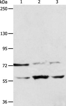
ApplicationsFlow Cytometry, ImmunoFluorescence, Western Blot, ELISA
Product group Antibodies
ReactivityCanine, Human, Mouse, Rat
TargetSYVN1
Overview
- SupplierEverest Biotech
- Product NameGoat anti-SYVN1
- Delivery Days Customer5
- Application Supplier NoteImmunofluorescence: Strong expression of the protein seen in the nuclei and endoplasmic reticulum of A431 cells. Recommended concentration: 10microg/ml. Flow Cytometry: Flow cytometric analysis of A431 cells. Recommended concentration: 10ug/ml.
- ApplicationsFlow Cytometry, ImmunoFluorescence, Western Blot, ELISA
- Applications SupplierPep-ELISA, WB, IF, FC
- CertificationResearch Use Only
- ClonalityPolyclonal
- Concentration0.5 mg/ml
- Gene ID84447
- Target nameSYVN1
- Target descriptionsynoviolin 1
- Target synonymsDER3, HRD1, E3 ubiquitin-protein ligase synoviolin, HMG-coA reductase degradation 1 homolog, RING-type E3 ubiquitin transferase synoviolin, synovial apoptosis inhibitor 1, synoviolin
- HostGoat
- Scientific DescriptionRefSeq number(s): NP_115807.1; NP_757385.1. GeneIDs all Nonhuman: 361712 (rat);. Purification: Antigen affinity purified. Names and symbols: SYVN1; synovial apoptosis inhibitor 1, synoviolin; HRD1; KIAA1810; MGC40372; synoviolin 1
- ReactivityCanine, Human, Mouse, Rat
- Reactivity SupplierHuman, Mouse, Rat, Dog
- Storage Instruction-20°C
- UNSPSC12352203
Related products
Product group Antibodies
Anti-SYVN1 AntibodyA37839
ApplicationsWestern Blot, ImmunoHistoChemistry
ReactivityHuman
- SizePrice

Product group Antibodies
Anti-HRD1/SYVN1 Antibody Picoband(r)A02670-3-CARRIER-FREE
ApplicationsFlow Cytometry, ImmunoFluorescence, Western Blot, ELISA, ImmunoCytoChemistry
ReactivityHuman, Mouse, Rat
TargetSYVN1
- SizePrice
Product group Antibodies
Anti-SYVN1 Antibody144-02605
ApplicationsWestern Blot
ReactivityHuman, Mouse
TargetSYVN1
- SizePrice
Product group Antibodies

SYVN1 / HRD1 AntibodyLS-C668919
ApplicationsImmunoPrecipitation, Western Blot
ReactivityHuman, Mouse, Rat
TargetSYVN1
- SizePrice
Product group Antibodies
References
SYVN1 Polyclonal AntibodyBS-0679R
ApplicationsFlow Cytometry, ImmunoFluorescence, Western Blot, ELISA, ImmunoCytoChemistry, ImmunoHistoChemistry, ImmunoHistoChemistry Frozen, ImmunoHistoChemistry Paraffin
ReactivityBovine, Canine, Human, Mouse, Rat
TargetSYVN1
- SizePrice
Product group Antibodies

Goat anti-SYVN1EB09822
ApplicationsELISA, ImmunoHistoChemistry
ReactivityHuman, Rat
TargetSYVN1
- SizePrice
Product group Antibodies
SYVN1 AntibodyCSB-PA803115LA01HU
ApplicationsImmunoFluorescence, ELISA, ImmunoHistoChemistry
ReactivityHuman
TargetSYVN1
- SizePrice

Product group Antibodies
Anti-SYVN1 AntibodyHPA005480
ApplicationsImmunoCytoChemistry, ImmunoHistoChemistry
ReactivityHuman
TargetSYVN1
- SizePrice
Product group Antibodies
SYVN1 antibody [C3-2], C-termGTX118990
ApplicationsImmunoPrecipitation, Western Blot
ReactivityHuman
TargetSYVN1
- SizePrice
Product group Antibodies
Anti-SYVN1 AntibodyCAB2605
ApplicationsWestern Blot, ELISA
ReactivityHuman
TargetSYVN1
- SizePrice