

SYVN1 antibody [C3-2], C-term
GTX118990
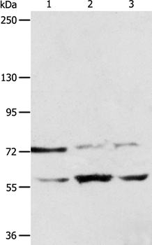
ApplicationsImmunoPrecipitation, Western Blot
Product group Antibodies
ReactivityHuman
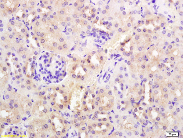
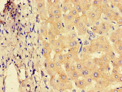
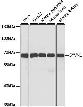
TargetSYVN1
Overview
- SupplierGeneTex
- Product NameSYVN1 antibody [C3-2], C-term
- Delivery Days Customer9
- ApplicationsImmunoPrecipitation, Western Blot
- CertificationResearch Use Only
- ClonalityPolyclonal
- Concentration1 mg/ml
- ConjugateUnconjugated
- Gene ID84447
- Target nameSYVN1
- Target descriptionsynoviolin 1
- Target synonymsDER3, HRD1, E3 ubiquitin-protein ligase synoviolin, HMG-coA reductase degradation 1 homolog, RING-type E3 ubiquitin transferase synoviolin, synovial apoptosis inhibitor 1, synoviolin
- HostRabbit
- IsotypeIgG
- Protein IDQ86TM6
- Protein NameE3 ubiquitin-protein ligase synoviolin
- Scientific DescriptionThis gene encodes a protein involved in endoplasmic reticulum (ER)-associated degradation. The encoded protein removes unfolded proteins, accumulated during ER stress, by retrograde transport to the cytosol from the ER. This protein also uses the ubiquitin-proteasome system for additional degradation of unfolded proteins. This gene and the mitochondrial ribosomal protein L49 gene use in their respective 3 UTRs some of the same genomic sequence. Sequence analysis identified two transcript variants that encode different isoforms. [provided by RefSeq]
- ReactivityHuman
- Storage Instruction-20°C or -80°C,2°C to 8°C
- UNSPSC41116161
Datasheet
Related products
Product group Antibodies
Anti-SYVN1 AntibodyA37839
ApplicationsWestern Blot, ImmunoHistoChemistry
ReactivityHuman
- SizePrice

Product group Antibodies
Anti-HRD1/SYVN1 Antibody Picoband(r)A02670-3-CARRIER-FREE
ApplicationsFlow Cytometry, ImmunoFluorescence, Western Blot, ELISA, ImmunoCytoChemistry
ReactivityHuman, Mouse, Rat
TargetSYVN1
- SizePrice
Product group Antibodies
Anti-SYVN1 Antibody144-02605
ApplicationsWestern Blot
ReactivityHuman, Mouse
TargetSYVN1
- SizePrice
Product group Antibodies

SYVN1 / HRD1 AntibodyLS-C668919
ApplicationsImmunoPrecipitation, Western Blot
ReactivityHuman, Mouse, Rat
TargetSYVN1
- SizePrice
Product group Antibodies
References
SYVN1 Polyclonal AntibodyBS-0679R
ApplicationsFlow Cytometry, ImmunoFluorescence, Western Blot, ELISA, ImmunoCytoChemistry, ImmunoHistoChemistry, ImmunoHistoChemistry Frozen, ImmunoHistoChemistry Paraffin
ReactivityBovine, Canine, Human, Mouse, Rat
TargetSYVN1
- SizePrice
Product group Antibodies

Goat anti-SYVN1EB09821
ApplicationsFlow Cytometry, ImmunoFluorescence, Western Blot, ELISA
ReactivityCanine, Human, Mouse, Rat
TargetSYVN1
- SizePrice
Product group Antibodies
SYVN1 AntibodyCSB-PA803115LA01HU
ApplicationsImmunoFluorescence, ELISA, ImmunoHistoChemistry
ReactivityHuman
TargetSYVN1
- SizePrice

Product group Antibodies
Anti-SYVN1 AntibodyHPA005480
ApplicationsImmunoCytoChemistry, ImmunoHistoChemistry
ReactivityHuman
TargetSYVN1
- SizePrice
Product group Antibodies
SYVN1 antibodyGTX66037
ApplicationsWestern Blot
ReactivityHuman, Mouse
TargetSYVN1
- SizePrice
Product group Antibodies
Anti-SYVN1 AntibodyCAB2605
ApplicationsWestern Blot, ELISA
ReactivityHuman
TargetSYVN1
- SizePrice