


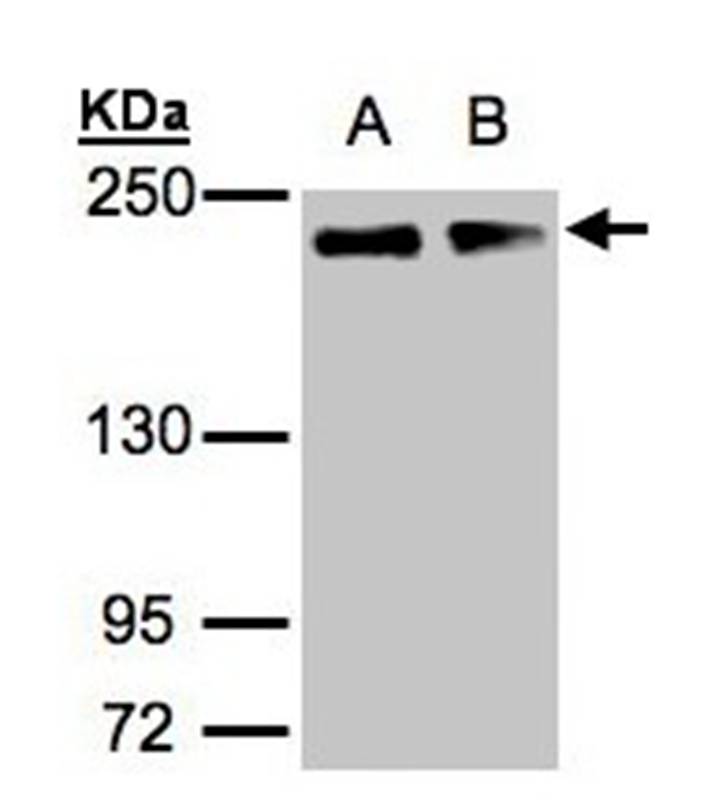
WB analysis of HeLa lysate with (B) and without (A) blocking with the immunising peptide using GTX89474 GOLGA3 antibody, C-term. Dilution : 0.1μg/ml Loading : 35μg protein in RIPA buffer
WB analysis of HeLa lysate with (B) and without (A) blocking with the immunising peptide using GTX89474 GOLGA3 antibody, C-term. Dilution : 0.1μg/ml Loading : 35μg protein in RIPA buffer
GOLGA3 antibody, C-term
GTX89474
ApplicationsWestern Blot
Product group Antibodies
ReactivityHuman
TargetGOLGA3
Overview
- SupplierGeneTex
- Product NameGOLGA3 antibody, C-term
- Delivery Days Customer9
- Application Supplier NoteWB: 0.1-0.3microg/ml. *Optimal dilutions/concentrations should be determined by the researcher.Not tested in other applications.
- ApplicationsWestern Blot
- CertificationResearch Use Only
- ClonalityPolyclonal
- Concentration0.50 mg/ml
- ConjugateUnconjugated
- Gene ID2802
- Target nameGOLGA3
- Target descriptiongolgin A3
- Target synonymsGCP170, MEA-2, golgin subfamily A member 3, Golgi membrane associated protein, Golgi peripheral membrane protein, SY2/SY10 protein, golgi autoantigen, golgin subfamily a, 3, golgi complex-associated protein of 170 kDa, golgin-160, golgin-165, male enhanced antigen-2
- HostGoat
- IsotypeIgG
- Protein IDQ08378
- Protein NameGolgin subfamily A member 3
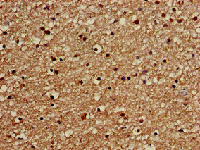
- Scientific DescriptionThe Golgi apparatus, which participates in glycosylation and transport of proteins and lipids in the secretory pathway, consists of a series of stacked cisternae (flattened membrane sacs). Interactions between the Golgi and microtubules are thought to be important for the reorganization of the Golgi after it fragments during mitosis. This gene encodes a member of the golgin family of proteins which are localized to the Golgi. Its encoded protein has been postulated to play a role in nuclear transport and Golgi apparatus localization. Several alternatively spliced transcript variants that encode different protein isoforms have been described for this gene. [provided by RefSeq, Feb 2010]
- ReactivityHuman
- Storage Instruction-20°C or -80°C,2°C to 8°C
- UNSPSC41116161
Datasheet
Related products
Product group Antibodies
GOLGA3 AntibodyCSB-PA600096LA01HU
ApplicationsImmunoFluorescence, ELISA, ImmunoHistoChemistry
ReactivityHuman
TargetGOLGA3
- SizePrice

Product group Antibodies
Anti-GOLGA3 Antibody Picoband(r)A07522-2-CARRIER-FREE
ApplicationsImmunoFluorescence, Western Blot, ELISA, ImmunoCytoChemistry, ImmunoHistoChemistry
ReactivityHuman
TargetGOLGA3
- SizePrice
Product group Antibodies
Anti-GOLGA3 AntibodyA34898
ApplicationsImmunoFluorescence, Western Blot, ImmunoHistoChemistry
ReactivityHuman
- SizePrice
Product group Antibodies

Goat anti-GOLGA3EB06926
ApplicationsWestern Blot, ELISA
ReactivityHuman
TargetGOLGA3
- SizePrice
Product group Antibodies
Anti-GOLGA3 AntibodyHPA039809
ApplicationsWestern Blot, ImmunoCytoChemistry, ImmunoHistoChemistry
ReactivityHuman
TargetGOLGA3
- SizePrice
Product group Antibodies

GOLGA3 Antibody (Biotin)LS-C500654
ApplicationsELISA
ReactivityHuman
TargetGOLGA3
- SizePrice
![Whole cell extract (30 μg) was separated by 5% SDS-PAGE, and the membrane was blotted with GOLGA3 antibody [C3], C-term (GTX100288) diluted at 1:1000. The HRP-conjugated anti-rabbit IgG antibody (GTX213110-01) was used to detect the primary antibody, and the signal was developed with Trident ECL plus-Enhanced.](https://www.genetex.com/upload/website/prouct_img/normal/GTX100288/GTX100288_40268_20220617_WB_22062121_743.webp)
Product group Antibodies
GOLGA3 antibody [C3], C-termGTX100288
ApplicationsImmunoFluorescence, Western Blot, ImmunoCytoChemistry, ImmunoHistoChemistry, ImmunoHistoChemistry Paraffin
ReactivityHuman
TargetGOLGA3
- SizePrice
Product group Antibodies
Anti-GOLGA3 (C-term) Antibody107-10205
ApplicationsImmunoFluorescence, Western Blot, ImmunoCytoChemistry, ImmunoHistoChemistry, ImmunoHistoChemistry Paraffin
ReactivityHuman
TargetGOLGA3
- SizePrice
Product group Antibodies
GOLGA3 Polyclonal AntibodyBS-13480R
ApplicationsImmunoFluorescence, Western Blot, ELISA, ImmunoCytoChemistry, ImmunoHistoChemistry, ImmunoHistoChemistry Frozen, ImmunoHistoChemistry Paraffin
ReactivityBovine, Human, Mouse, Rabbit, Rat, Sheep
TargetGOLGA3
- SizePrice