

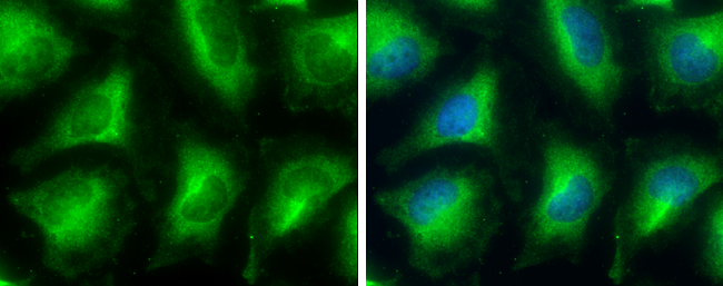
GPD1 antibody detects GPD1 protein at cytoplasm by immunofluorescent analysis. Sample: HeLa cells were fixed in 4% paraformaldehyde at RT for 15 min. Green: GPD1 protein stained by GPD1 antibody (GTX117910) diluted at 1:500. Blue: Hoechst 33342 staining.
 dilution: 1:500.
Antigen Retrieval: Trilogy? (EDTA based, pH 8.0) buffer, 15min")
 A: A549 10% SDS PAGE GTX117910 diluted at 1:1000")
 antibody at 1:500 dilution.
Antigen Retrieval: Trilogy? (EDTA based, pH 8.0) buffer, 15min")
 dilution: 1:500.
Antigen Retrieval: Trilogy? (EDTA based, pH 8.0) buffer, 15min")
GPD1 antibody detects GPD1 protein at cytoplasm by immunofluorescent analysis. Sample: HeLa cells were fixed in 4% paraformaldehyde at RT for 15 min. Green: GPD1 protein stained by GPD1 antibody (GTX117910) diluted at 1:500. Blue: Hoechst 33342 staining.
GPD1 antibody
GTX117910
ApplicationsImmunoFluorescence, Western Blot, ImmunoCytoChemistry, ImmunoHistoChemistry, ImmunoHistoChemistry Paraffin
Product group Antibodies
ReactivityHuman, Rat
TargetGPD1
Overview
- SupplierGeneTex
- Product NameGPD1 antibody
- Delivery Days Customer9
- Application Supplier NoteWB: 1:500-1:3000. ICC/IF: 1:100-1:1000. IHC-P: 1:100-1:1000. *Optimal dilutions/concentrations should be determined by the researcher.Not tested in other applications.
- ApplicationsImmunoFluorescence, Western Blot, ImmunoCytoChemistry, ImmunoHistoChemistry, ImmunoHistoChemistry Paraffin
- CertificationResearch Use Only
- ClonalityPolyclonal
- Concentration0.51 mg/ml
- ConjugateUnconjugated
- Gene ID2819
- Target nameGPD1
- Target descriptionglycerol-3-phosphate dehydrogenase 1
- Target synonymsGPD-C, GPDH-C, HTGTI, glycerol-3-phosphate dehydrogenase [NAD(+)], cytoplasmic, epididymis secretory sperm binding protein, glycerol-3-phosphate dehydrogenase 1 (soluble), glycerol-3-phosphate dehydrogenase [NAD+], cytoplasmic, glycerophosphate dehydrogenase
- HostRabbit
- IsotypeIgG
- Protein IDP21695
- Protein NameGlycerol-3-phosphate dehydrogenase [NAD(+)], cytoplasmic
- ReactivityHuman, Rat
- Storage Instruction-20°C or -80°C,2°C to 8°C
- UNSPSC41116161
Datasheet
Related products
Product group Antibodies
Anti-GPD1 AntibodyA31001
ApplicationsWestern Blot, ImmunoHistoChemistry
ReactivityHuman, Mouse
- SizePrice
Product group Antibodies
Anti-GPD1 Antibody144-05715
ApplicationsWestern Blot, ImmunoHistoChemistry
ReactivityHuman, Mouse, Rat
TargetGPD1
- SizePrice
Product group Antibodies
GPD1 Polyclonal AntibodyBS-2388R
ApplicationsImmunoFluorescence, Western Blot, ELISA, ImmunoCytoChemistry, ImmunoHistoChemistry, ImmunoHistoChemistry Frozen, ImmunoHistoChemistry Paraffin
ReactivityBovine, Equine, Human, Mouse, Porcine, Rabbit, Rat
TargetGPD1
- SizePrice
Product group Antibodies
GPD1 AntibodyCSB-PA009709HA01HU
ApplicationsImmunoFluorescence, ELISA, ImmunoHistoChemistry
ReactivityHuman
TargetGPD1
- SizePrice
Product group Antibodies

GPD1 AntibodyLS-C334237
ApplicationsWestern Blot, ImmunoHistoChemistry
ReactivityHuman, Mouse, Rat
TargetGPD1
- SizePrice

Product group Antibodies
Anti-GPD1 AntibodyHPA058621
ApplicationsImmunoHistoChemistry
ReactivityHuman
TargetGPD1
- SizePrice
![Non-transfected (–) and transfected (+) 293T whole cell extracts (30 μg) were separated by 10% SDS-PAGE, and the membrane was blotted with GPD1 antibody [HL1959] (GTX637796) diluted at 1:5000. The HRP-conjugated anti-rabbit IgG antibody (GTX213110-01) was used to detect the primary antibody.](https://www.genetex.com/upload/website/prouct_img/normal/GTX637796/GTX637796_T-44851_20230428_WB_multiple_B_23050223_653.webp)
Product group Antibodies
GPD1 antibody [HL1959]GTX637796
ApplicationsImmunoFluorescence, Western Blot, ImmunoCytoChemistry, ImmunoHistoChemistry, ImmunoHistoChemistry Paraffin
ReactivityHuman, Mouse, Rat
TargetGPD1
- SizePrice
Product group Antibodies
GPD1 antibodyGTX32630
ApplicationsWestern Blot, ImmunoHistoChemistry, ImmunoHistoChemistry Paraffin
ReactivityHuman, Mouse, Rat
TargetGPD1
- SizePrice
Product group Antibodies
Anti-GPD1 AntibodyCAB5715
ApplicationsWestern Blot, ELISA
ReactivityHuman
TargetGPD1
- SizePrice