

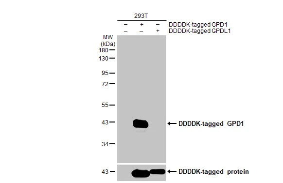
Non-transfected (–) and transfected (+) 293T whole cell extracts (30 μg) were separated by 10% SDS-PAGE, and the membrane was blotted with GPD1 antibody [HL1959] (GTX637796) diluted at 1:5000. The HRP-conjugated anti-rabbit IgG antibody (GTX213110-01) was used to detect the primary antibody.
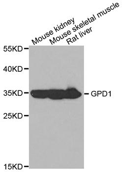
![Various tissue extracts (50 μg) were separated by 10% SDS-PAGE, and the membrane was blotted with GPD1 antibody [HL1959] (GTX637796) diluted at 1:5000. The HRP-conjugated anti-rabbit IgG antibody (GTX213110-01) was used to detect the primary antibody.](https://www.genetex.com/upload/website/prouct_img/normal/GTX637796/GTX637796_T-44851_20230512_WB_M_R_23051702_830.webp "Various tissue extracts (50 μg) were separated by 10% SDS-PAGE, and the membrane was blotted with GPD1 antibody [HL1959] (GTX637796) diluted at 1:5000. The HRP-conjugated anti-rabbit IgG antibody (GTX213110-01) was used to detect the primary antibody.")
![Various whole cell extracts (30 μg) were separated by 10% SDS-PAGE, and the membrane was blotted with GPD1 antibody [HL1959] (GTX637796) diluted at 1:1000. The HRP-conjugated anti-rabbit IgG antibody (GTX213110-01) was used to detect the primary antibody. Corresponding RNA expression data for the same cell lines are based on Human Protein Atlas program.](https://www.genetex.com/upload/website/prouct_img/normal/GTX637796/GTX637796_45089_20230707_WB_TPM_watermark_23071223_702.webp "Various whole cell extracts (30 μg) were separated by 10% SDS-PAGE, and the membrane was blotted with GPD1 antibody [HL1959] (GTX637796) diluted at 1:1000. The HRP-conjugated anti-rabbit IgG antibody (GTX213110-01) was used to detect the primary antibody. Corresponding RNA expression data for the same cell lines are based on Human Protein Atlas program.")
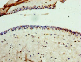
![GPD1 antibody [HL1959] detects GPD1 protein at cytoplasm by immunohistochemical analysis. Sample: Paraffin-embedded mouse liver. GPD1 stained by GPD1 antibody [HL1959] (GTX637796) diluted at 1:100. Antigen Retrieval: Citrate buffer, pH 6.0, 15 min](https://www.genetex.com/upload/website/prouct_img/normal/GTX637796/GTX637796_T-44851_20230801_IHC-P_M_23081619_919.webp "GPD1 antibody [HL1959] detects GPD1 protein at cytoplasm by immunohistochemical analysis. Sample: Paraffin-embedded mouse liver. GPD1 stained by GPD1 antibody [HL1959] (GTX637796) diluted at 1:100. Antigen Retrieval: Citrate buffer, pH 6.0, 15 min")
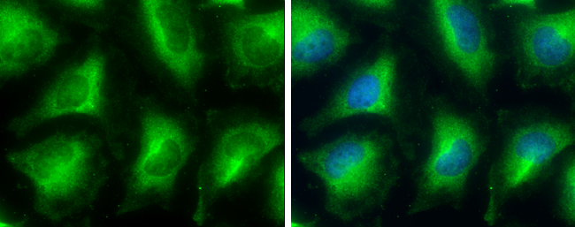
![GPD1 antibody [HL1959] detects GPD1 protein at cell membrane and cytoplasm by immunofluorescent analysis. Sample: Caco-2 cells were fixed in ice-cold MeOH for 5 min. Green: GPD1 stained by GPD1 antibody [HL1959] (GTX637796) diluted at 1:500. Blue: Fluoroshield with DAPI (GTX30920).](https://www.genetex.com/upload/website/prouct_img/normal/GTX637796/GTX637796_45089_20231006_ICC_IF_23102401_538.webp "GPD1 antibody [HL1959] detects GPD1 protein at cell membrane and cytoplasm by immunofluorescent analysis. Sample: Caco-2 cells were fixed in ice-cold MeOH for 5 min. Green: GPD1 stained by GPD1 antibody [HL1959] (GTX637796) diluted at 1:500. Blue: Fluoroshield with DAPI (GTX30920).")
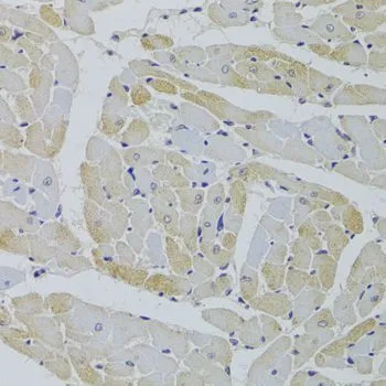
![GPD1 antibody [HL1959] detects GPD1 protein by immunohistochemical analysis. Sample: Paraffin-embedded human tissues. GPD1 stained by GPD1 antibody [HL1959] (GTX637796) diluted at 1:100. Antigen Retrieval: Citrate buffer, pH 6.0, 15 min](https://www.genetex.com/upload/website/prouct_img/normal/GTX637796/GTX637796_45089_20240923_IHC-P_multiple_24092600_859.webp "GPD1 antibody [HL1959] detects GPD1 protein by immunohistochemical analysis. Sample: Paraffin-embedded human tissues. GPD1 stained by GPD1 antibody [HL1959] (GTX637796) diluted at 1:100. Antigen Retrieval: Citrate buffer, pH 6.0, 15 min")
Non-transfected (–) and transfected (+) 293T whole cell extracts (30 μg) were separated by 10% SDS-PAGE, and the membrane was blotted with GPD1 antibody [HL1959] (GTX637796) diluted at 1:5000. The HRP-conjugated anti-rabbit IgG antibody (GTX213110-01) was used to detect the primary antibody.
GPD1 antibody [HL1959]
GTX637796
ApplicationsImmunoFluorescence, Western Blot, ImmunoCytoChemistry, ImmunoHistoChemistry, ImmunoHistoChemistry Paraffin
Product group Antibodies
ReactivityHuman, Mouse, Rat
TargetGPD1
Overview
- SupplierGeneTex
- Product NameGPD1 antibody [HL1959]
- Delivery Days Customer9
- Application Supplier NoteWB: 1:500-1:10000. *Optimal dilutions/concentrations should be determined by the researcher.Not tested in other applications.
- ApplicationsImmunoFluorescence, Western Blot, ImmunoCytoChemistry, ImmunoHistoChemistry, ImmunoHistoChemistry Paraffin
- CertificationResearch Use Only
- ClonalityMonoclonal
- Clone IDHL1959
- Concentration1 mg/ml
- ConjugateUnconjugated
- Gene ID2819
- Target nameGPD1
- Target descriptionglycerol-3-phosphate dehydrogenase 1
- Target synonymsGPD-C, GPDH-C, HTGTI, glycerol-3-phosphate dehydrogenase [NAD(+)], cytoplasmic, epididymis secretory sperm binding protein, glycerol-3-phosphate dehydrogenase 1 (soluble), glycerol-3-phosphate dehydrogenase [NAD+], cytoplasmic, glycerophosphate dehydrogenase
- HostRabbit
- IsotypeIgG
- Protein IDP21695
- Protein NameGlycerol-3-phosphate dehydrogenase [NAD(+)], cytoplasmic
- Scientific DescriptionThis gene encodes a member of the NAD-dependent glycerol-3-phosphate dehydrogenase family. The encoded protein plays a critical role in carbohydrate and lipid metabolism by catalyzing the reversible conversion of dihydroxyacetone phosphate (DHAP) and reduced nicotine adenine dinucleotide (NADH) to glycerol-3-phosphate (G3P) and NAD+. The encoded cytosolic protein and mitochondrial glycerol-3-phosphate dehydrogenase also form a glycerol phosphate shuttle that facilitates the transfer of reducing equivalents from the cytosol to mitochondria. Mutations in this gene are a cause of transient infantile hypertriglyceridemia. Alternatively spliced transcript variants encoding multiple isoforms have been observed for this gene. [provided by RefSeq, Mar 2012]
- ReactivityHuman, Mouse, Rat
- Storage Instruction-20°C or -80°C,2°C to 8°C
- UNSPSC41116161
Datasheet
Related products
Product group Antibodies
Anti-GPD1 AntibodyA31001
ApplicationsWestern Blot, ImmunoHistoChemistry
ReactivityHuman, Mouse
- SizePrice
Product group Antibodies
Anti-GPD1 Antibody144-05715
ApplicationsWestern Blot, ImmunoHistoChemistry
ReactivityHuman, Mouse, Rat
TargetGPD1
- SizePrice
Product group Antibodies
GPD1 Polyclonal AntibodyBS-2388R
ApplicationsImmunoFluorescence, Western Blot, ELISA, ImmunoCytoChemistry, ImmunoHistoChemistry, ImmunoHistoChemistry Frozen, ImmunoHistoChemistry Paraffin
ReactivityBovine, Equine, Human, Mouse, Porcine, Rabbit, Rat
TargetGPD1
- SizePrice
Product group Antibodies
GPD1 AntibodyCSB-PA009709HA01HU
ApplicationsImmunoFluorescence, ELISA, ImmunoHistoChemistry
ReactivityHuman
TargetGPD1
- SizePrice
Product group Antibodies

GPD1 AntibodyLS-C334237
ApplicationsWestern Blot, ImmunoHistoChemistry
ReactivityHuman, Mouse, Rat
TargetGPD1
- SizePrice

Product group Antibodies
Anti-GPD1 AntibodyHPA058621
ApplicationsImmunoHistoChemistry
ReactivityHuman
TargetGPD1
- SizePrice
Product group Antibodies
GPD1 antibodyGTX117910
ApplicationsImmunoFluorescence, Western Blot, ImmunoCytoChemistry, ImmunoHistoChemistry, ImmunoHistoChemistry Paraffin
ReactivityHuman, Rat
TargetGPD1
- SizePrice
Product group Antibodies
GPD1 antibodyGTX32630
ApplicationsWestern Blot, ImmunoHistoChemistry, ImmunoHistoChemistry Paraffin
ReactivityHuman, Mouse, Rat
TargetGPD1
- SizePrice
Product group Antibodies
Anti-GPD1 AntibodyCAB5715
ApplicationsWestern Blot, ELISA
ReactivityHuman
TargetGPD1
- SizePrice