

Growth Associated Protein 43 (GAP43) Polyclonal Antibody
CAU26135
ApplicationsImmunoPrecipitation, Western Blot, ImmunoCytoChemistry, ImmunoHistoChemistry
Product group Antibodies
ReactivityMouse
TargetGap43
Overview
- SupplierBiomatik
- Product NameGrowth Associated Protein 43 (GAP43) Polyclonal Antibody
- Delivery Days Customer12
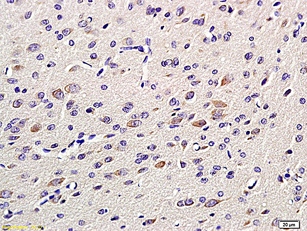
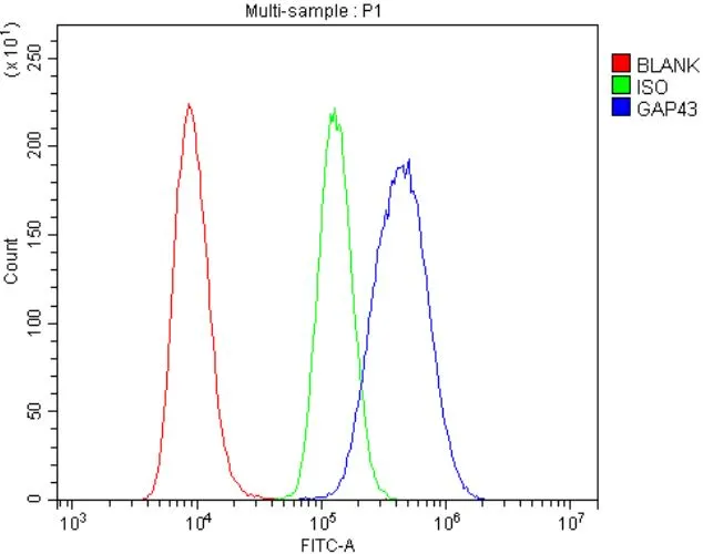
- ApplicationsImmunoPrecipitation, Western Blot, ImmunoCytoChemistry, ImmunoHistoChemistry
- Applications SupplierWB; IHC; ICC; IP.
- CertificationResearch Use Only
- ClonalityPolyclonal
- Concentration0.5 mg/ml
- ConjugateUnconjugated
- Gene ID14432
- Target nameGap43
- Target descriptiongrowth associated protein 43
- Target synonymsB-50, Basp2, GAP-43, neuromodulin, axonal membrane protein GAP-43, brain abundant, membrane attached signal protein 2, calmodulin-binding protein P-57, growth accentuating protein 43
- HostRabbit
- Protein IDP06837
- Protein NameNeuromodulin
- Scientific DescriptionThe Growth Associated Protein 43 (GAP43) Polyclonal Antibody (Species: Mouse) has been validated for the following applications: WB, IHC, ICC, IP.
- ReactivityMouse
- Reactivity SupplierMouse
- Storage Instruction-20°C,2°C to 8°C
- UNSPSC12352203
Related products
Product group Antibodies
References
GAP43 Polyclonal AntibodyBS-0154R
ApplicationsFlow Cytometry, ImmunoFluorescence, Western Blot, ELISA, ImmunoCytoChemistry, ImmunoHistoChemistry, ImmunoHistoChemistry Frozen, ImmunoHistoChemistry Paraffin
ReactivityCanine, Chicken, Human, Mouse, Rat
TargetGap43
- SizePrice
Product group Antibodies
GAP43 antibodyGTX04779
ApplicationsFlow Cytometry, Western Blot, ELISA, ImmunoHistoChemistry, ImmunoHistoChemistry Paraffin
ReactivityHuman, Mouse, Rat
TargetGap43
- SizePrice

Product group Antibodies
Anti-GAP43 Antibody Picoband(r)A01868-1-CARRIER-FREE
ApplicationsFlow Cytometry, Western Blot, ELISA, ImmunoHistoChemistry
ReactivityHuman, Mouse, Rat
TargetGap43
- SizePrice