

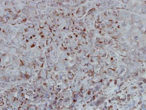
Immunohistochemical analysis of paraffin-embedded H661 xenograft, using GSK3 beta(GTX111192) antibody at 1:500 dilution.
Antigen Retrieval: Trilogy? (EDTA based, pH 8.0) buffer, 15min
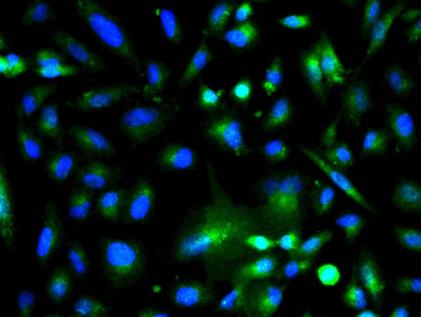
![GSK3 beta antibody [C1C3] detects GSK3 beta protein at cytoplasm and cell membrane by immunofluorescent analysis. Sample: HeLa cells were fixed in 4% paraformaldehyde at RT for 15 min. Green: GSK3 beta stained by GSK3 beta antibody [C1C3] (GTX111192) diluted at 1:500. Blue: Fluoroshield with DAPI (GTX30920). Scale bar= 10μm.](https://www.genetex.com/upload/website/prouct_img/normal/GTX111192/GTX111192_44384_20220408_ICC_IF_w_23060500_975.webp "GSK3 beta antibody [C1C3] detects GSK3 beta protein at cytoplasm and cell membrane by immunofluorescent analysis. Sample: HeLa cells were fixed in 4% paraformaldehyde at RT for 15 min. Green: GSK3 beta stained by GSK3 beta antibody [C1C3] (GTX111192) diluted at 1:500. Blue: Fluoroshield with DAPI (GTX30920). Scale bar= 10μm.")
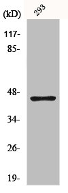
![Non-transfected (–) and transfected (+) 293T whole cell extracts (30 μg) were separated by 10% SDS-PAGE, and the membrane was blotted with GSK3 beta antibody [C1C3] (GTX111192) diluted at 1:1000. The HRP-conjugated anti-rabbit IgG antibody (GTX213110-01) was used to detect the primary antibody.](https://www.genetex.com/upload/website/prouct_img/normal/GTX111192/GTX111192_40051_20160526_WB_shRNA_watermark_w_23060500_458.webp "Non-transfected (–) and transfected (+) 293T whole cell extracts (30 μg) were separated by 10% SDS-PAGE, and the membrane was blotted with GSK3 beta antibody [C1C3] (GTX111192) diluted at 1:1000. The HRP-conjugated anti-rabbit IgG antibody (GTX213110-01) was used to detect the primary antibody.")
 A: mouse brain 10% SDS PAGE GTX111192 diluted at 1:3000 The HRP-conjugated anti-rabbit IgG antibody (GTX213110-01) was used to detect the primary antibody.")
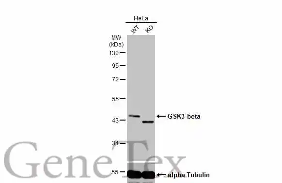
![Whole cell extract (30 μg) was separated by 10% SDS-PAGE, and the membrane was blotted with GSK3 beta antibody [C1C3] (GTX111192) diluted at 1:500. The HRP-conjugated anti-rabbit IgG antibody (GTX213110-01) was used to detect the primary antibody.](https://www.genetex.com/upload/website/prouct_img/normal/GTX111192/GTX111192_40051_20170811_WB_w_23060500_559.webp "Whole cell extract (30 μg) was separated by 10% SDS-PAGE, and the membrane was blotted with GSK3 beta antibody [C1C3] (GTX111192) diluted at 1:500. The HRP-conjugated anti-rabbit IgG antibody (GTX213110-01) was used to detect the primary antibody.")
![GSK3 beta antibody [C1C3] detects GSK3 beta protein at cytoplasm by immunofluorescent analysis. Sample: SK-N-SH cells were fixed in 4% paraformaldehyde at RT for 15 min. Green: GSK3 beta protein stained by GSK3 beta antibody [C1C3] (GTX111192) diluted at 1:400. Blue: Hoechst 33342 staining. Scale bar = 10 μm.](https://www.genetex.com/upload/website/prouct_img/normal/GTX111192/GTX111192_40051_20160325_IFA_w_23060500_994.webp "GSK3 beta antibody [C1C3] detects GSK3 beta protein at cytoplasm by immunofluorescent analysis. Sample: SK-N-SH cells were fixed in 4% paraformaldehyde at RT for 15 min. Green: GSK3 beta protein stained by GSK3 beta antibody [C1C3] (GTX111192) diluted at 1:400. Blue: Hoechst 33342 staining. Scale bar = 10 μm.")
![GSK3 beta antibody [C1C3] detects GSK3 beta protein at cytoplasm in rat brain by immunohistochemical analysis. Sample: Paraffin-embedded rat brain. GSK3 beta antibody [C1C3] (GTX111192) diluted at 1:500.
Antigen Retrieval: Citrate buffer, pH 6.0, 15 min](https://www.genetex.com/upload/website/prouct_img/normal/GTX111192/GTX111192_40051_20160127_IHC-P_R_w_23060500_291.webp "GSK3 beta antibody [C1C3] detects GSK3 beta protein at cytoplasm in rat brain by immunohistochemical analysis. Sample: Paraffin-embedded rat brain. GSK3 beta antibody [C1C3] (GTX111192) diluted at 1:500.
Antigen Retrieval: Citrate buffer, pH 6.0, 15 min")

Immunohistochemical analysis of paraffin-embedded H661 xenograft, using GSK3 beta(GTX111192) antibody at 1:500 dilution.
Antigen Retrieval: Trilogy? (EDTA based, pH 8.0) buffer, 15min
GSK3 beta antibody [C1C3]
GTX111192
ApplicationsImmunoFluorescence, Western Blot, ImmunoCytoChemistry, ImmunoHistoChemistry, ImmunoHistoChemistry Paraffin
Product group Antibodies
ReactivityHuman, Mouse, Rat
TargetGSK3B
Overview
- SupplierGeneTex
- Product NameGSK3 beta antibody [C1C3]
- Delivery Days Customer9
- Application Supplier NoteWB: 1:500-1:3000. ICC/IF: 1:100-1:1000. IHC-P: 1:100-1:1000. *Optimal dilutions/concentrations should be determined by the researcher.Not tested in other applications.
- ApplicationsImmunoFluorescence, Western Blot, ImmunoCytoChemistry, ImmunoHistoChemistry, ImmunoHistoChemistry Paraffin
- CertificationResearch Use Only
- ClonalityPolyclonal
- Concentration1.57 mg/ml
- ConjugateUnconjugated
- Gene ID2932
- Target nameGSK3B
- Target descriptionglycogen synthase kinase 3 beta
- Target synonymsglycogen synthase kinase-3 beta, GSK-3 beta, serine/threonine-protein kinase GSK3B
- HostRabbit
- IsotypeIgG
- Protein IDP49841
- Protein NameGlycogen synthase kinase-3 beta
- Scientific DescriptionGlycogen synthase kinase-3 (GSK3) is a proline-directed serine-threonine kinase that was initially identified as a phosphorylating and inactivating glycogen synthase (see GYS1, MIM 138570). Two isoforms, alpha (GSK3A; MIM 606784) and beta, show a high degree of amino acid homology (Stambolic and Woodgett, 1994 [PubMed 7980435]). GSK3B is involved in energy metabolism, neuronal cell development, and body pattern formation (Plyte et al., 1992 [PubMed 1333807]).[supplied by OMIM]
- ReactivityHuman, Mouse, Rat
- Storage Instruction-20°C or -80°C,2°C to 8°C
- UNSPSC41116161
Datasheet
Related products
Product group Antibodies
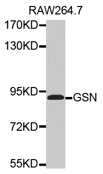
Anti-GSN AntibodyA30655
ApplicationsWestern Blot, ImmunoHistoChemistry
ReactivityHuman, Mouse, Rat
- SizePrice

Product group Antibodies
Anti-GSK3 beta/GSK3B Antibody Picoband(r)A00791-3-CARRIER-FREE
ApplicationsImmunoFluorescence, Western Blot, ELISA, ImmunoCytoChemistry, ImmunoHistoChemistry
ReactivityHuman, Mouse, Rat
TargetGSK3B
- SizePrice
Product group Antibodies
ApplicationsELISA
ReactivityHuman
TargetGSK3B
- SizePrice
Product group Antibodies

GSK3B / GSK3 Beta Antibody (clone 4C3)LS-C767028
ApplicationsWestern Blot, ImmunoHistoChemistry, ImmunoHistoChemistry Paraffin
ReactivityHuman, Mouse, Rat
TargetGSK3B
- SizePrice
Product group Antibodies
References
GSK-3 Beta Polyclonal AntibodyBS-0028R
ApplicationsFlow Cytometry, ImmunoFluorescence, Western Blot, ELISA, ImmunoCytoChemistry, ImmunoHistoChemistry, ImmunoHistoChemistry Frozen, ImmunoHistoChemistry Paraffin
ReactivityHuman, Mouse, Rat
TargetGSK3B
- SizePrice
Product group Antibodies
GSK3B AntibodyCSB-PA002848
ApplicationsImmunoPrecipitation, Western Blot, ELISA, ImmunoHistoChemistry
ReactivityHuman, Mouse, Rat
TargetGSK3B
- SizePrice
Product group Antibodies
GSK3B Polyclonal AntibodyCAC13750
ApplicationsImmunoFluorescence, Western Blot, ELISA
ReactivityMouse
TargetGSK3B
- SizePrice
Product group Antibodies
ApplicationsWestern Blot, ImmunoHistoChemistry, ImmunoHistoChemistry Frozen
ReactivityHuman, Mouse
TargetGSK3B
- SizePrice
Product group Antibodies
GSK3 beta antibodyGTX133372
ApplicationsImmunoFluorescence, Western Blot, ImmunoCytoChemistry
ReactivityHuman, Mouse, Rat
TargetGSK3B
- SizePrice