

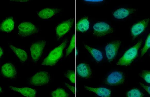
GSTP1 antibody detects GSTP1 protein at cytoplasm and nucleus by immunofluorescent analysis. Sample: HeLa cells were fixed in 4% paraformaldehyde at RT for 15 min. Green: GSTP1 stained by GSTP1 antibody (GTX112953) diluted at 1:500. Blue: Fluoroshield with DAPI (GTX30920).
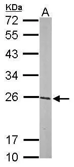
 A: mouse brain 12% SDS PAGE GTX112953 diluted at 1:1000 The HRP-conjugated anti-rabbit IgG antibody (GTX213110-01) was used to detect the primary antibody.")
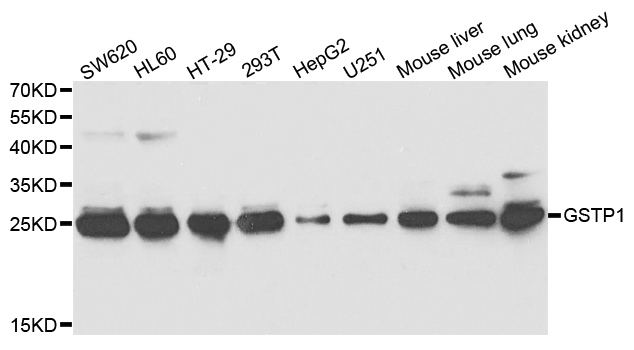
 dilution: 1:1000 The HRP-conjugated anti-rabbit IgG antibody (GTX213110-01) was used to detect the primary antibody.")
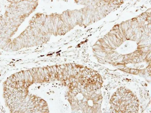
 diluted at 1:500. Antigen Retrieval: Citrate buffer, pH 6.0, 15 min")
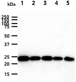
 were separated by 12% SDS-PAGE, and the membrane was blotted with GSTP1 antibody (GTX112953) diluted at 1:1000. The HRP-conjugated anti-rabbit IgG antibody (GTX213110-01) was used to detect the primary antibody. Corresponding RNA expression data for the same cell lines are based on Human Protein Atlas program.")

GSTP1 antibody detects GSTP1 protein at cytoplasm and nucleus by immunofluorescent analysis. Sample: HeLa cells were fixed in 4% paraformaldehyde at RT for 15 min. Green: GSTP1 stained by GSTP1 antibody (GTX112953) diluted at 1:500. Blue: Fluoroshield with DAPI (GTX30920).
GSTP1 antibody
GTX112953
ApplicationsImmunoFluorescence, Western Blot, ImmunoCytoChemistry, ImmunoHistoChemistry, ImmunoHistoChemistry Paraffin
Product group Antibodies
ReactivityHuman, Mouse, Rat
TargetGSTP1
Overview
- SupplierGeneTex
- Product NameGSTP1 antibody
- Delivery Days Customer9
- Application Supplier NoteWB: 1:500-1:3000. ICC/IF: 1:100-1:1000. IHC-P: 1:100-1:1000. *Optimal dilutions/concentrations should be determined by the researcher.Not tested in other applications.
- ApplicationsImmunoFluorescence, Western Blot, ImmunoCytoChemistry, ImmunoHistoChemistry, ImmunoHistoChemistry Paraffin
- CertificationResearch Use Only
- ClonalityPolyclonal
- Concentration0.92 mg/ml
- ConjugateUnconjugated
- Gene ID2950
- Target nameGSTP1
- Target descriptionglutathione S-transferase pi 1
- Target synonymsDFN7, FAEES3, GST3, GSTP, HEL-S-22, PI, glutathione S-transferase P, GST class-pi, GSTP1-1, deafness, X-linked 7, epididymis secretory protein Li 22, fatty acid ethyl ester synthase III
- HostRabbit
- IsotypeIgG
- Protein IDP09211
- Protein NameGlutathione S-transferase P
- Scientific DescriptionGlutathione S-transferases (GSTs) are a family of enzymes that play an important role in detoxification by catalyzing the conjugation of many hydrophobic and electrophilic compounds with reduced glutathione. Based on their biochemical, immunologic, and structural properties, the soluble GSTs are categorized into 4 main classes: alpha, mu, pi, and theta. This GST family member is a polymorphic gene encoding active, functionally different GSTP1 variant proteins that are thought to function in xenobiotic metabolism and play a role in susceptibility to cancer, and other diseases. [provided by RefSeq]
- ReactivityHuman, Mouse, Rat
- Storage Instruction-20°C or -80°C,2°C to 8°C
- UNSPSC12352203
References
- Xu YM, Tan HW, Zheng W, et al. Cadmium telluride quantum dot-exposed human bronchial epithelial cells: a further study of the cellular response by proteomics. Toxicol Res (Camb). 2019,8(6):994-1001. doi: 10.1039/c9tx00126cRead this paper
- Chen DJ, Xu YM, Zheng W, et al. Proteomic analysis of secreted proteins by human bronchial epithelial cells in response to cadmium toxicity. Proteomics. 2015,15(17):3075-86. doi: 10.1002/pmic.201400489Read this paper
- Hsiao CY, Hung CY, Tsai TH, et al. A Study of the Wound Healing Mechanism of a Traditional Chinese Medicine, Angelica sinensis, Using a Proteomic Approach. Evid Based Complement Alternat Med. 2012,2012:467531. doi: 10.1155/2012/467531Read this paper
Datasheet
Related products
Product group Antibodies

Anti-GSTP1 [SAIC-22D-22]AB00312-1.1-BT
ApplicationsMass Spectrometry
ReactivityHuman
TargetGSTP1
- SizePrice
Product group Antibodies
Anti-GSTP1 Antibody130-10294
ApplicationsWestern Blot, ELISA
ReactivityHuman
- SizePrice
Product group Antibodies
References
GSTP1 antibody [N1N2], N-termGTX100299
ApplicationsImmunoFluorescence, Western Blot, ImmunoCytoChemistry, ImmunoHistoChemistry, ImmunoHistoChemistry Paraffin
ReactivityHuman, Mouse
TargetGSTP1
- SizePrice
Product group Antibodies
References
GSTP1 antibodyGTX112695
ApplicationsImmunoFluorescence, Western Blot, ImmunoCytoChemistry, ImmunoHistoChemistry, ImmunoHistoChemistry Frozen, ImmunoHistoChemistry Paraffin
ReactivityHuman, Mouse, Rat
TargetGSTP1
- SizePrice
Product group Antibodies
GSTP1 antibody [AT12C10]GTX57728
ApplicationsFlow Cytometry, ImmunoFluorescence, Western Blot, ImmunoCytoChemistry
ReactivityHuman
TargetGSTP1
- SizePrice
Product group Antibodies
Anti-GSTP1 AntibodyA30827
ApplicationsFlow Cytometry, ImmunoFluorescence, ImmunoPrecipitation, Western Blot, ImmunoHistoChemistry
ReactivityHuman, Mouse, Rat
- SizePrice
![IHC-P analysis of human prostate tissue using GTX83077 GSTP1 antibody [3F2C2].](https://www.genetex.com/upload/website/prouct_img/normal/GTX83077/GTX83077_20170912_IHC-P_w_23061322_450.webp)
Product group Antibodies
GSTP1 antibody [3F2C2]GTX83077
ApplicationsFlow Cytometry, ImmunoFluorescence, Western Blot, ELISA, ImmunoCytoChemistry, ImmunoHistoChemistry, ImmunoHistoChemistry Paraffin
ReactivityHuman
TargetGSTP1
- SizePrice
Product group Antibodies
GSTP1 Polyclonal AntibodyCAC14121
ApplicationsWestern Blot, ELISA
TargetGSTP1
- SizePrice
Product group Antibodies
GSTP1 Recombinant AntibodyBSM-60581R
ApplicationsFlow Cytometry, ImmunoFluorescence, Western Blot, ImmunoCytoChemistry, ImmunoHistoChemistry, ImmunoHistoChemistry Frozen, ImmunoHistoChemistry Paraffin
ReactivityHuman
TargetGSTP1
- SizePrice