

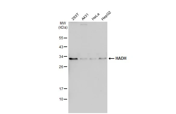
Various whole cell extracts (30 μg) were separated by 12% SDS-PAGE, and the membrane was blotted with HADH antibody (GTX118325) diluted at 1:1000. The HRP-conjugated anti-rabbit IgG antibody (GTX213110-01) was used to detect the primary antibody.
![HADH antibody detects HADH protein at mitochondria by immunofluorescent analysis. Sample: HepG2 cells were fixed in 4% paraformaldehyde at RT for 15 min. Green: HADH stained by HADH antibody (GTX118325) diluted at 1:500. Red: alpha Tubulin, a cytoskeleton marker, stained by alpha Tubulin antibody [GT114] (GTX628802) diluted at 1:1000. Blue: Fluoroshield with DAPI (GTX30920).](https://www.genetex.com/upload/website/prouct_img/normal/GTX118325/GTX118325_44104_20211224_ICC_IF_w_23060519_335.webp "HADH antibody detects HADH protein at mitochondria by immunofluorescent analysis. Sample: HepG2 cells were fixed in 4% paraformaldehyde at RT for 15 min. Green: HADH stained by HADH antibody (GTX118325) diluted at 1:500. Red: alpha Tubulin, a cytoskeleton marker, stained by alpha Tubulin antibody [GT114] (GTX628802) diluted at 1:1000. Blue: Fluoroshield with DAPI (GTX30920).")
 was separated by 12% SDS-PAGE, and the membrane was blotted with HADH antibody (GTX118325) diluted at 1:1000. The HRP-conjugated anti-rabbit IgG antibody (GTX213110-01) was used to detect the primary antibody.")
 dilution: 1:250.
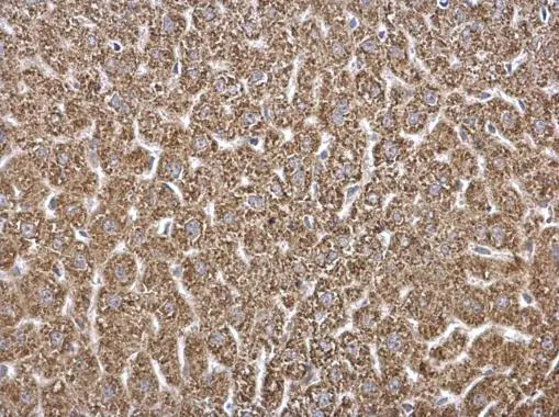
Antigen Retrieval: Trilogy? (EDTA based, pH 8.0) buffer, 15min")
 was separated by 12% SDS-PAGE, and the membrane was blotted with HADH antibody (GTX118325) diluted at 1:1000. The HRP-conjugated anti-rabbit IgG antibody (GTX213110-01) was used to detect the primary antibody.")
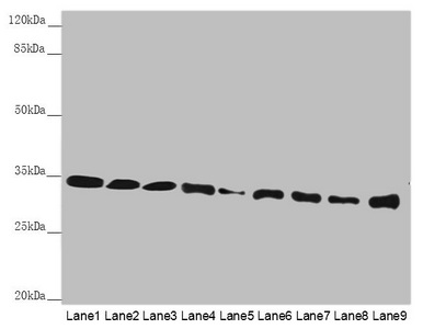
 A: mouse liver 10% SDS PAGE GTX118325 diluted at 1:1000 The HRP-conjugated anti-rabbit IgG antibody (GTX213110-01) was used to detect the primary antibody.")
 diluted at 1:500. Blue: Hoechst 33343 staining.")
Various whole cell extracts (30 μg) were separated by 12% SDS-PAGE, and the membrane was blotted with HADH antibody (GTX118325) diluted at 1:1000. The HRP-conjugated anti-rabbit IgG antibody (GTX213110-01) was used to detect the primary antibody.
HADH antibody
GTX118325
ApplicationsImmunoFluorescence, Western Blot, ImmunoCytoChemistry, ImmunoHistoChemistry, ImmunoHistoChemistry Paraffin
Product group Antibodies
ReactivityHuman, Mouse, Rat
TargetHADH
Overview
- SupplierGeneTex
- Product NameHADH antibody
- Delivery Days Customer9
- Application Supplier NoteWB: 1:500-1:3000. ICC/IF: 1:100-1:1000. IHC-P: 1:100-1:1000. *Optimal dilutions/concentrations should be determined by the researcher.Not tested in other applications.
- ApplicationsImmunoFluorescence, Western Blot, ImmunoCytoChemistry, ImmunoHistoChemistry, ImmunoHistoChemistry Paraffin
- CertificationResearch Use Only
- ClonalityPolyclonal
- Concentration0.49 mg/ml
- ConjugateUnconjugated
- Gene ID3033
- Target nameHADH
- Target descriptionhydroxyacyl-CoA dehydrogenase
- Target synonymsHAD, HADH1, HADHSC, HCDH, HHF4, MSCHAD, SCHAD, hydroxyacyl-coenzyme A dehydrogenase, mitochondrial, L-3-hydroxyacyl-Coenzyme A dehydrogenase, short chain, medium and short-chain L-3-hydroxyacyl-coenzyme A dehydrogenase, short-chain 3-hydroxyacyl-CoA dehydrogenase, testis secretory sperm-binding protein Li 203a
- HostRabbit
- IsotypeIgG
- Protein IDQ16836
- Protein NameHydroxyacyl-coenzyme A dehydrogenase, mitochondrial
- Scientific DescriptionThis gene is a member of the 3-hydroxyacyl-CoA dehydrogenase gene family. The encoded protein functions in the mitochondrial matrix to catalyze the oxidation of straight-chain 3-hydroxyacyl-CoAs as part of the beta-oxidation pathway. Its enzymatic activity is highest with medium-chain-length fatty acids. Mutations in this gene cause one form of familial hyperinsulinemic hypoglycemia. The human genome contains a related pseudogene. [provided by RefSeq]
- ReactivityHuman, Mouse, Rat
- Storage Instruction-20°C or -80°C,2°C to 8°C
- UNSPSC41116161
Datasheet
Related products
Product group Antibodies
ApplicationsImmunoPrecipitation, Western Blot, ImmunoCytoChemistry, ImmunoHistoChemistry
ReactivityPorcine
TargetHADH
- SizePrice
Product group Antibodies
HADH AntibodyCSB-PA614992ESR1HU
ApplicationsWestern Blot, ELISA, ImmunoHistoChemistry
ReactivityHuman, Mouse
TargetHADH
- SizePrice
Product group Antibodies
Anti-HADH AntibodyA116629
ApplicationsDot Blot, ImmunoFluorescence, Western Blot, ELISA
ReactivityPorcine
- SizePrice
Product group Antibodies
Anti-HADH Antibody144-01076
ApplicationsWestern Blot, ImmunoHistoChemistry
ReactivityHuman, Mouse, Rat
TargetHADH
- SizePrice
Product group Antibodies

Goat anti-HADH / HADHSCEB08200
ApplicationsWestern Blot, ELISA
ReactivityCanine, Human, Mouse, Rat
TargetHADH
- SizePrice

Product group Antibodies
Anti-HADH AntibodyHPA039588
ApplicationsWestern Blot, ImmunoHistoChemistry
ReactivityHuman
TargetHADH
- SizePrice

Product group Antibodies
Anti-HADH Antibody Picoband(r)A03650-1-CARRIER-FREE
ApplicationsFlow Cytometry, ImmunoFluorescence, Western Blot, ELISA, ImmunoCytoChemistry, ImmunoHistoChemistry
ReactivityHuman, Mouse, Rat
TargetHADH
- SizePrice
Product group Antibodies

HADH AntibodyLS-C331243
ApplicationsWestern Blot, ImmunoHistoChemistry
ReactivityHuman, Mouse, Rat
TargetHADH
- SizePrice
Product group Antibodies
HADH antibodyGTX105167
ApplicationsImmunoFluorescence, ImmunoPrecipitation, Western Blot, ImmunoCytoChemistry, ImmunoHistoChemistry, ImmunoHistoChemistry Paraffin
ReactivityHuman, Mouse, Rat
TargetHADH
- SizePrice