

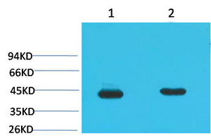
HEK293T cells were transfected with the pCMV6-ENTRY control (Left lane) or pCMV6-ENTRY HAO1 (RC216834, Right lane) cDNA for 48 hrs and lysed. Equivalent amounts of cell lysates (5 ug per lane) were separated by SDS-PAGE and immunoblotted with anti-HAO1. Positive lysates LY413692 (100ug) and LC413692 (20ug) can be purchased separately from OriGene.
HEK293T cells were transfected with the pCMV6-ENTRY control (Left lane) or pCMV6-ENTRY HAO1 (RC216834, Right lane) cDNA for 48 hrs and lysed. Equivalent amounts of cell lysates (5 ug per lane) were separated by SDS-PAGE and immunoblotted with anti-HAO1. Positive lysates LY413692 (100ug) and LC413692 (20ug) can be purchased separately from OriGene.
HAO1 Mouse Monoclonal Antibody [Clone ID: OTI3E7]
CF501937
ApplicationsFlow Cytometry, Western Blot
Product group Antibodies
ReactivityHuman, Mouse, Rat
TargetHAO1
Overview
- SupplierOriGene
- Product NameHAO1 Mouse Monoclonal Antibody [Clone ID: OTI3E7]
- Delivery Days Customer14
- ApplicationsFlow Cytometry, Western Blot
- CertificationResearch Use Only
- ClonalityMonoclonal
- Clone IDOTI3E7
- Gene ID54363
- Target nameHAO1
- Target descriptionhydroxyacid oxidase 1
- Target synonymsGO, GOX, GOX1, HAOX1, 2-Hydroxyacid oxidase 1, (S)-2-hydroxy-acid oxidase, glycolate oxidase 1, glyoxylate oxidase, hydroxyacid oxidase (glycolate oxidase) 1
- HostMouse
- IsotypeIgG1
- Protein IDQ9UJM8
- Protein Name2-Hydroxyacid oxidase 1
- Scientific DescriptionCarrier-free (BSA/glycerol-free) HAO1 mouse monoclonal antibody, clone OTI3E7 (formerly 3E7)
- ReactivityHuman, Mouse, Rat
- Storage Instruction-20°C
- UNSPSC12352203
MSDS
Related products

Product group Antibodies
Anti-HAO1 Antibody Picoband(r)A09159-2-CARRIER-FREE
ApplicationsFlow Cytometry, Western Blot, ELISA, ImmunoHistoChemistry
ReactivityHuman, Mouse, Rat
TargetHAO1
- SizePrice
Product group Antibodies
Anti-HAO1 AntibodyA41277
ApplicationsWestern Blot
ReactivityMouse, Rat
- SizePrice
Product group Antibodies

HAO1 Antibody (clone 3B2)LS-C767091
ApplicationsImmunoFluorescence, Western Blot, ImmunoHistoChemistry, ImmunoHistoChemistry Paraffin
ReactivityMouse, Rat
TargetHAO1
- SizePrice

Product group Antibodies
Anti-HAO1 AntibodyHPA049552
ApplicationsWestern Blot, ImmunoHistoChemistry
ReactivityHuman
TargetHAO1
- SizePrice
Product group Antibodies
HAO1 Monoclonal AntibodyCSB-MA080172
ApplicationsWestern Blot, ELISA
ReactivityMouse, Rat
TargetHAO1
- SizePrice
![IHC-P analysis of human colon tissue using GTX33987 HAO1 antibody [Mix]. Negative control (the lower left coner) was secondary antibody only. Antigen retrieval : Sodium citrate pH6.0 was used for antibody retrieval (>98oC, 20min) Dilution : 1:200](https://www.genetex.com/upload/website/prouct_img/normal/GTX33987/GTX33987_20200622_IHC-P_064_w_23060801_325.webp)
Product group Antibodies
HAO1 antibody [Mix]GTX33987
ApplicationsWestern Blot, ImmunoHistoChemistry, ImmunoHistoChemistry Paraffin
ReactivityHuman, Mouse, Rat
TargetHAO1
- SizePrice
Product group Antibodies
Anti-HAO1 Antibody144-06470
ApplicationsImmunoFluorescence, Western Blot, ImmunoHistoChemistry
ReactivityHuman, Mouse, Rat
TargetHAO1
- SizePrice
Product group Antibodies
HAO1 Polyclonal AntibodyBS-8602R
ApplicationsImmunoFluorescence, Western Blot, ELISA, ImmunoCytoChemistry, ImmunoHistoChemistry, ImmunoHistoChemistry Frozen, ImmunoHistoChemistry Paraffin
ReactivityCanine, Equine, Human, Mouse, Porcine, Rabbit, Rat
- SizePrice