

Hdac1 Polyclonal Antibody
CAC07026
ApplicationsImmunoFluorescence, ChIP Chromatin ImmunoPrecipitation, ELISA, ImmunoHistoChemistry
Product group Antibodies
TargetHDAC1
Overview
- SupplierBiomatik
- Product NameHdac1 Polyclonal Antibody
- Delivery Days Customer12
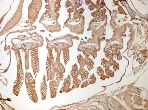
- ApplicationsImmunoFluorescence, ChIP Chromatin ImmunoPrecipitation, ELISA, ImmunoHistoChemistry
- Applications SupplierELISA, IHC, IF, ChIP; Recommended dilution: IHC:1:20-1:200, IF:1:100-1:500
- CertificationResearch Use Only
- ClonalityPolyclonal
- ConjugateUnconjugated
- Gene ID3065
- Target nameHDAC1
- Target descriptionhistone deacetylase 1
- Target synonymsGON-10, HD1, KDAC1, RPD3, RPD3L1, histone deacetylase 1, protein deacetylase HDAC1, protein deacylase HDAC1, protein decrotonylase HDAC1, reduced potassium dependency, yeast homolog-like 1
- HostRabbit
- IsotypeIgG
- Protein IDQ13547
- Protein NameHistone deacetylase 1
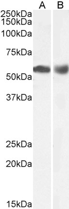
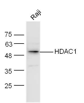
- Scientific DescriptionThe Hdac1 Polyclonal Antibody (Species: Human) has been validated for the following applications: ELISA, IHC, IF, ChIP.
- Reactivity SupplierHuman
- Storage Instruction-20°C,2°C to 8°C
- UNSPSC12352203
Related products
Product group Antibodies
Anti-HDAC1 AntibodyA84192
ApplicationsImmunoFluorescence, Western Blot, ChIP Chromatin ImmunoPrecipitation, ELISA
ReactivityHuman, Mouse
- SizePrice
Product group Antibodies

ApplicationsImmunoFluorescence, Western Blot, ELISA, ImmunoCytoChemistry, ImmunoHistoChemistry
- SizePrice
Product group Antibodies
Anti-HDAC1 Antibody144-00238
ApplicationsImmunoFluorescence, ImmunoPrecipitation, Western Blot, ImmunoHistoChemistry
ReactivityHuman, Mouse, Rat
TargetHDAC1
- SizePrice

Product group Antibodies
Anti-HDAC1 AntibodyAMAB90781
ApplicationsWestern Blot, ImmunoCytoChemistry, ImmunoHistoChemistry
ReactivityHuman, Mouse, Rat
TargetHDAC1
- SizePrice

Product group Antibodies
Anti-HDAC1 Antibody Picoband(r)A00256-4-CARRIER-FREE
ApplicationsFlow Cytometry, Western Blot, ImmunoHistoChemistry
ReactivityHuman
TargetHDAC1
- SizePrice

Product group Antibodies
HDAC1 AntibodyCSB-PA002877
ApplicationsWestern Blot, ELISA, ImmunoHistoChemistry
ReactivityHuman, Mouse, Rat
TargetHDAC1
- SizePrice
Product group Antibodies

ApplicationsImmunoFluorescence, Western Blot, ChIP Chromatin ImmunoPrecipitation, ELISA
ReactivityCanine, Human, Mouse, Rat
TargetHDAC1
- SizePrice
Product group Antibodies
References
HDAC1 Polyclonal AntibodyBS-1414R
ApplicationsFlow Cytometry, ImmunoFluorescence, Western Blot, ELISA, ImmunoCytoChemistry, ImmunoHistoChemistry, ImmunoHistoChemistry Frozen, ImmunoHistoChemistry Paraffin
ReactivityBovine, Canine, Guinea Pig, Human, Mouse, Porcine, Rat, Sheep
TargetHDAC1
- SizePrice
Product group Antibodies
HDAC1 antibodyGTX100513
ApplicationsImmunoFluorescence, ImmunoPrecipitation, Western Blot, ChIP Chromatin ImmunoPrecipitation, ImmunoCytoChemistry, ImmunoHistoChemistry, ImmunoHistoChemistry Frozen, ImmunoHistoChemistry Paraffin
ReactivityHuman, Mouse, Rat, Zebra Fish
TargetHDAC1
- SizePrice