

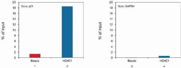
ChIP analysis of U-2OS cells using GTX20012 HDAC1 antibody. Left : ChIP results using the anti-HDAC1 antibody (bar 2) or beads only (bar 1) and PCR primers specific for p21 Right : ChIP results using the anti-HDAC1 antibody (bar 4) or beads only (bar 3) and PCR primers for GAPDH (used as negative control). ChIP reaction : 2.4microg antibody / 1 million cells
 or HeLa extract (Panel B) using GTX20012 HDAC1 antibody. WB was performed using anti- HA antibody or GTX20012 HDAC1 antibody as indicated. Lane 1 and 4 : Beads only Lane 2 and 5 : IP with 2 microg GTX20012 HDAC1 antibody Lane 3 : IP with HA antibody as a confrol")
 was performed to detect Xist RNA (green signal). Nuclei were DAPI-stained to specifically label the DNA. Fixation : formaldehyde Permeabilization : Triton X-100 Dilution : 1:200 and incubated for 45 minutes at room temperature")
, and the column flow through obtained from the antibody purification step. The antigen used was the C-terminal peptide used for immunization. By plotting the absorbance against the antibody dilution, the titer of the antibody was estimated to be 1:4,250.")
ChIP analysis of U-2OS cells using GTX20012 HDAC1 antibody. Left : ChIP results using the anti-HDAC1 antibody (bar 2) or beads only (bar 1) and PCR primers specific for p21 Right : ChIP results using the anti-HDAC1 antibody (bar 4) or beads only (bar 3) and PCR primers for GAPDH (used as negative control). ChIP reaction : 2.4microg antibody / 1 million cells
HDAC1 antibody
GTX20012
ApplicationsImmunoFluorescence, ImmunoPrecipitation, Western Blot, ChIP Chromatin ImmunoPrecipitation, ELISA, ImmunoCytoChemistry
Product group Antibodies
ReactivityHuman, Mouse
TargetHDAC1
Overview
- SupplierGeneTex
- Product NameHDAC1 antibody
- Delivery Days Customer9
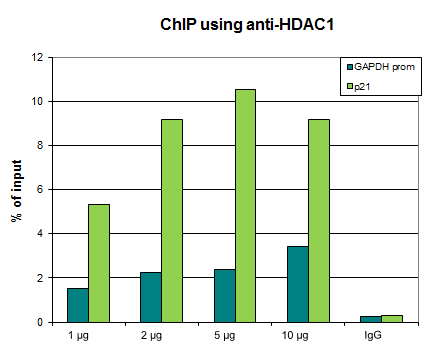
- Application Supplier NoteICC/IF: 1:200. ELISA: 1:100 - 1:300. ChIP assay: 2.4 microg. *Optimal dilutions/concentrations should be determined by the researcher.Not tested in other applications.
- ApplicationsImmunoFluorescence, ImmunoPrecipitation, Western Blot, ChIP Chromatin ImmunoPrecipitation, ELISA, ImmunoCytoChemistry
- CertificationResearch Use Only
- ClonalityPolyclonal
- ConjugateUnconjugated
- Gene ID3065
- Target nameHDAC1
- Target descriptionhistone deacetylase 1
- Target synonymsGON-10, HD1, KDAC1, RPD3, RPD3L1, histone deacetylase 1, protein deacetylase HDAC1, protein deacylase HDAC1, protein decrotonylase HDAC1, reduced potassium dependency, yeast homolog-like 1
- HostRabbit
- IsotypeIgG
- Protein IDQ13547
- Protein NameHistone deacetylase 1
- Scientific DescriptionHistone acetylation and deacetylation, catalyzed by multisubunit complexes, play a key role in the regulation of eukaryotic gene expression. The protein encoded by this gene belongs to the histone deacetylase/acuc/apha family and is a component of the histone deacetylase complex. It also interacts with retinoblastoma tumor-suppressor protein and this complex is a key element in the control of cell proliferation and differentiation. Together with metastasis-associated protein-2, it deacetylates p53 and modulates its effect on cell growth and apoptosis. [provided by RefSeq, Jul 2008]
- ReactivityHuman, Mouse
- Storage Instruction-20°C or -80°C,2°C to 8°C
- UNSPSC12352203
Datasheet
Related products
Product group Antibodies
Hdac1 Polyclonal AntibodyCAC07026
ApplicationsImmunoFluorescence, ChIP Chromatin ImmunoPrecipitation, ELISA, ImmunoHistoChemistry
TargetHDAC1
- SizePrice
Product group Antibodies
ApplicationsImmunoFluorescence, Western Blot, ChIP Chromatin ImmunoPrecipitation
ReactivityHuman
TargetHDAC1
- SizePrice
Product group Antibodies
Anti-HDAC1 AntibodyA84192
ApplicationsImmunoFluorescence, Western Blot, ChIP Chromatin ImmunoPrecipitation, ELISA
ReactivityHuman, Mouse
- SizePrice

Product group Antibodies
Anti-HDAC1 AntibodyAMAB90781
ApplicationsWestern Blot, ImmunoCytoChemistry, ImmunoHistoChemistry
ReactivityHuman, Mouse, Rat
TargetHDAC1
- SizePrice
Product group Antibodies
Anti-HDAC1 Antibody144-00238
ApplicationsImmunoFluorescence, ImmunoPrecipitation, Western Blot, ImmunoHistoChemistry
ReactivityHuman, Mouse, Rat
TargetHDAC1
- SizePrice
Product group Antibodies

ApplicationsImmunoFluorescence, Western Blot, ELISA, ImmunoCytoChemistry, ImmunoHistoChemistry
- SizePrice
Product group Antibodies

ApplicationsImmunoFluorescence, Western Blot, ChIP Chromatin ImmunoPrecipitation, ELISA
ReactivityCanine, Human, Mouse, Rat
TargetHDAC1
- SizePrice
Product group Antibodies
ApplicationsWestern Blot, ImmunoHistoChemistry, ImmunoHistoChemistry Paraffin
ReactivityHuman
TargetHDAC1
- SizePrice
Product group Antibodies
References
HDAC1 antibodyGTX27028
ApplicationsImmunoFluorescence, ImmunoPrecipitation, Western Blot, ImmunoCytoChemistry, ImmunoHistoChemistry, ImmunoHistoChemistry Paraffin
ReactivityHuman, Mouse, Rat
TargetHDAC1
- SizePrice