

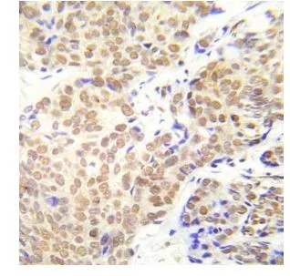
IHC-P analysis of human breast carcinoma tissue using GTX79062 HDAC1 (phospho Ser421/Ser423) antibody.
 antibody. Left : Primary antibody Right : Primary antibody preincubated with immunogen peptide")
IHC-P analysis of human breast carcinoma tissue using GTX79062 HDAC1 (phospho Ser421/Ser423) antibody.
HDAC1 (phospho Ser421/Ser423) antibody
GTX79062
ApplicationsWestern Blot, ImmunoHistoChemistry, ImmunoHistoChemistry Paraffin
Product group Antibodies
ReactivityHuman
TargetHDAC1
Overview
- SupplierGeneTex
- Product NameHDAC1 (phospho Ser421/Ser423) antibody
- Delivery Days Customer9
- Application Supplier NoteWB: 1:500 - 1:1000. IHC-P: 1:50 - 1:100. *Optimal dilutions/concentrations should be determined by the researcher.Not tested in other applications.
- ApplicationsWestern Blot, ImmunoHistoChemistry, ImmunoHistoChemistry Paraffin
- CertificationResearch Use Only
- ClonalityPolyclonal
- ConjugateUnconjugated
- Gene ID3065
- Target nameHDAC1
- Target descriptionhistone deacetylase 1
- Target synonymsGON-10, HD1, KDAC1, RPD3, RPD3L1, histone deacetylase 1, protein deacetylase HDAC1, protein deacylase HDAC1, protein decrotonylase HDAC1, reduced potassium dependency, yeast homolog-like 1
- HostRabbit
- IsotypeIgG
- Protein IDQ13547
- Protein NameHistone deacetylase 1
- Scientific DescriptionHistone acetylation and deacetylation, catalyzed by multisubunit complexes, play a key role in the regulation of eukaryotic gene expression. The protein encoded by this gene belongs to the histone deacetylase/acuc/apha family and is a component of the histone deacetylase complex. It also interacts with retinoblastoma tumor-suppressor protein and this complex is a key element in the control of cell proliferation and differentiation. Together with metastasis-associated protein-2, it deacetylates p53 and modulates its effect on cell growth and apoptosis. [provided by RefSeq, Jul 2008]
- ReactivityHuman
- Storage Instruction-20°C or -80°C,2°C to 8°C
- UNSPSC12352203
Datasheet
Related products
Product group Antibodies
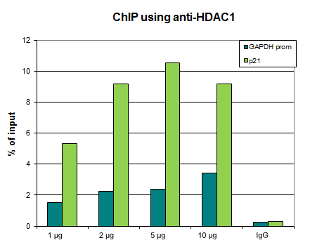
ApplicationsImmunoFluorescence, Western Blot, ChIP Chromatin ImmunoPrecipitation
ReactivityHuman
TargetHDAC1
- SizePrice
Product group Antibodies

ApplicationsImmunoFluorescence, Western Blot, ELISA, ImmunoCytoChemistry, ImmunoHistoChemistry
- SizePrice
Product group Antibodies
Anti-HDAC1 Antibody144-00238
ApplicationsImmunoFluorescence, ImmunoPrecipitation, Western Blot, ImmunoHistoChemistry
ReactivityHuman, Mouse, Rat
TargetHDAC1
- SizePrice

Product group Antibodies
Anti-HDAC1 Antibody Picoband(r)A00256-4-CARRIER-FREE
ApplicationsFlow Cytometry, Western Blot, ImmunoHistoChemistry
ReactivityHuman
TargetHDAC1
- SizePrice
![WB analysis of various samples using GTX08970 HDAC1 antibody [GT1163]. Dilution : 1:1000 Loading : 25μg](https://www.genetex.com/upload/website/prouct_img/normal/GTX08970/GTX08970_20200327_WB_1_w_23053123_389.webp)
Product group Antibodies
HDAC1 antibody [GT1163]GTX08970
ApplicationsImmunoFluorescence, Western Blot, ImmunoCytoChemistry, ImmunoHistoChemistry, ImmunoHistoChemistry Paraffin
ReactivityHuman, Mouse, Rat
TargetHDAC1
- SizePrice
Product group Antibodies
References
HDAC1 antibodyGTX100513
ApplicationsImmunoFluorescence, ImmunoPrecipitation, Western Blot, ChIP Chromatin ImmunoPrecipitation, ImmunoCytoChemistry, ImmunoHistoChemistry, ImmunoHistoChemistry Frozen, ImmunoHistoChemistry Paraffin
ReactivityHuman, Mouse, Rat, Zebra Fish
TargetHDAC1
- SizePrice
![Wild-type (WT) and HDAC1 knockout (KO) HeLa cell extracts (30 μg) were separated by 10% SDS-PAGE, and the membrane was blotted with HDAC1 antibody [HL1691] (GTX637290) diluted at 1:1000. The HRP-conjugated anti-rabbit IgG antibody (GTX213110-01) was used to detect the primary antibody.](https://www.genetex.com/upload/website/prouct_img/normal/GTX637290/GTX637290_T-44767_20220902_WB_KO_watermark_22090701_939.webp)
Product group Antibodies
HDAC1 antibody [HL1691]GTX637290
ApplicationsImmunoFluorescence, Western Blot, ImmunoCytoChemistry
ReactivityHuman
TargetHDAC1
- SizePrice
Product group Antibodies
Anti-HDAC1Y058101
ApplicationsWestern Blot, ELISA
ReactivityHuman, Mouse
- SizePrice
Product group Antibodies
Hdac1 Polyclonal AntibodyCAC07026
ApplicationsImmunoFluorescence, ChIP Chromatin ImmunoPrecipitation, ELISA, ImmunoHistoChemistry
TargetHDAC1
- SizePrice