

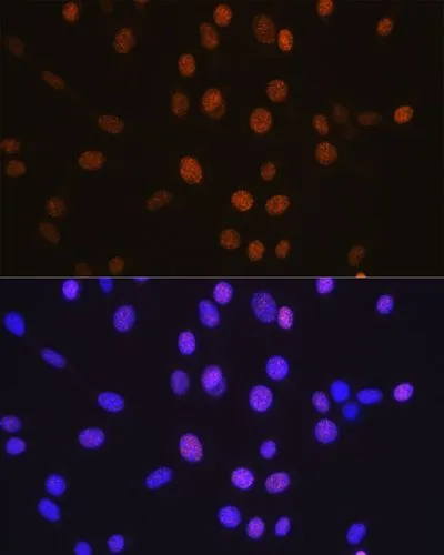
ICC/IF analysis of NIH-3T3 cells using GTX01527 HDAC2 antibody [GT1219]. Blue : DAPI
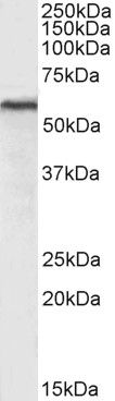
![WB analysis of various samples using GTX01527 HDAC2 antibody [GT1219]. Dilution : 1:1000 Loading : 25 μg](https://www.genetex.com/upload/website/prouct_img/normal/GTX01527/GTX01527_20200508_WB_w_23053121_202.webp "WB analysis of various samples using GTX01527 HDAC2 antibody [GT1219]. Dilution : 1:1000 Loading : 25 μg")
![IHC-P analysis of mouse brain tissue section using GTX01527 HDAC2 antibody [GT1219]. Dilution : 1:100](https://www.genetex.com/upload/website/prouct_img/normal/GTX01527/GTX01527_20200508_IHC-P_w_23053121_565.webp "IHC-P analysis of mouse brain tissue section using GTX01527 HDAC2 antibody [GT1219]. Dilution : 1:100")
![IHC-P analysis of rat spleen tissue section using GTX01527 HDAC2 antibody [GT1219]. Dilution : 1:100](https://www.genetex.com/upload/website/prouct_img/normal/GTX01527/GTX01527_20200508_IHC-P_2_w_23053121_451.webp "IHC-P analysis of rat spleen tissue section using GTX01527 HDAC2 antibody [GT1219]. Dilution : 1:100")
![ICC/IF analysis of C6 cells using GTX01527 HDAC2 antibody [GT1219]. Blue : DAPI](https://www.genetex.com/upload/website/prouct_img/normal/GTX01527/GTX01527_20200508_ICCIF_1_w_23053121_594.webp "ICC/IF analysis of C6 cells using GTX01527 HDAC2 antibody [GT1219]. Blue : DAPI")
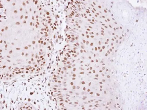
![IHC-P analysis of human oophoroma tissue section using GTX01527 HDAC2 antibody [GT1219]. Dilution : 1:100](https://www.genetex.com/upload/website/prouct_img/normal/GTX01527/GTX01527_20200508_IHC-P_1_w_23053121_184.webp "IHC-P analysis of human oophoroma tissue section using GTX01527 HDAC2 antibody [GT1219]. Dilution : 1:100")
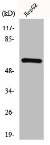
![Various whole cell extracts (30 μg) were separated by 10% SDS-PAGE, and the membrane was blotted with HDAC2 antibody [GT1219] (GTX01527) diluted at 1:1000. The HRP-conjugated anti-rabbit IgG antibody (GTX213110-01) was used to detect the primary antibody.](https://www.genetex.com/upload/website/prouct_img/normal/GTX01527/GTX01527_4000000105_20200410_WB_w_23053121_435.webp "Various whole cell extracts (30 μg) were separated by 10% SDS-PAGE, and the membrane was blotted with HDAC2 antibody [GT1219] (GTX01527) diluted at 1:1000. The HRP-conjugated anti-rabbit IgG antibody (GTX213110-01) was used to detect the primary antibody.")
ICC/IF analysis of NIH-3T3 cells using GTX01527 HDAC2 antibody [GT1219]. Blue : DAPI
HDAC2 antibody [GT1219]
GTX01527
ApplicationsImmunoFluorescence, Western Blot, ImmunoCytoChemistry, ImmunoHistoChemistry, ImmunoHistoChemistry Paraffin
Product group Antibodies
ReactivityHuman, Mouse, Rat
TargetHDAC2
Overview
- SupplierGeneTex
- Product NameHDAC2 antibody [GT1219]
- Delivery Days Customer9
- Application Supplier NoteWB: 1:500 - 1:2000. ICC/IF: 1:50 - 1:200. IHC-P: 1:50 - 1:200. *Optimal dilutions/concentrations should be determined by the researcher.Not tested in other applications.
- ApplicationsImmunoFluorescence, Western Blot, ImmunoCytoChemistry, ImmunoHistoChemistry, ImmunoHistoChemistry Paraffin
- CertificationResearch Use Only
- ClonalityMonoclonal
- Clone IDGT1219
- ConjugateUnconjugated
- Gene ID3066
- Target nameHDAC2
- Target descriptionhistone deacetylase 2
- Target synonymsHD2, KDAC2, RPD3, YAF1, histone deacetylase 2, YY1-associated factor 1, protein deacylase HDAC2, transcriptional regulator homolog RPD3
- HostRabbit
- IsotypeIgG
- Protein IDQ92769
- Protein NameHistone deacetylase 2
- Scientific DescriptionThis gene product belongs to the histone deacetylase family. Histone deacetylases act via the formation of large multiprotein complexes, and are responsible for the deacetylation of lysine residues at the N-terminal regions of core histones (H2A, H2B, H3 and H4). This protein forms transcriptional repressor complexes by associating with many different proteins, including YY1, a mammalian zinc-finger transcription factor. Thus, it plays an important role in transcriptional regulation, cell cycle progression and developmental events. Alternative splicing results in multiple transcript variants. [provided by RefSeq, Apr 2010]
- ReactivityHuman, Mouse, Rat
- Storage Instruction-20°C or -80°C,2°C to 8°C
- UNSPSC41116161
References
- Protein phosphatase 2A inactivation induces microsatellite instability, neoantigen production and immune response. Yen YT et al., 2021 Dec 15, Nat CommunRead this paper
Datasheet
Related products
Product group Antibodies
Anti-HDAC2 AntibodyA286069
ApplicationsFlow Cytometry, ImmunoFluorescence, Western Blot, ELISA
ReactivityHuman, Mouse
- SizePrice
Product group Antibodies
Anti-HDAC2 Antibody144-02084
ApplicationsImmunoFluorescence, ImmunoPrecipitation, Western Blot, ImmunoHistoChemistry
ReactivityHuman, Monkey, Mouse, Rat
TargetHDAC2
- SizePrice
Product group Antibodies

HDAC2 AntibodyLS-C761136
ApplicationsWestern Blot
ReactivityChicken, Human, Mouse, Rat
TargetHDAC2
- SizePrice

Product group Antibodies
Anti-HDAC2 Antibody Picoband(r)A00325-3-CARRIER-FREE
ApplicationsFlow Cytometry, ImmunoFluorescence, Western Blot, ELISA, ImmunoCytoChemistry, ImmunoHistoChemistry
ReactivityHuman, Mouse, Rat
TargetHDAC2
- SizePrice
Product group Antibodies
ApplicationsFlow Cytometry, ImmunoFluorescence, Western Blot, ImmunoCytoChemistry, ImmunoHistoChemistry, ImmunoHistoChemistry Frozen, ImmunoHistoChemistry Paraffin
ReactivityBovine, Canine, Chicken, Equine, Human, Mouse, Porcine, Rat
TargetHDAC2
- SizePrice
Product group Antibodies
HDAC2 AntibodyCSB-PA002879
ApplicationsWestern Blot, ELISA, ImmunoHistoChemistry
ReactivityHuman, Monkey, Mouse, Rat
TargetHDAC2
- SizePrice
Product group Antibodies

Goat anti-Hdac2 (mouse)EB11448
ApplicationsFlow Cytometry, ImmunoFluorescence, Western Blot, ELISA
ReactivityBovine, Canine, Human, Mouse, Porcine, Rat
TargetHDAC2
- SizePrice
Product group Antibodies
ApplicationsImmunoPrecipitation, Western Blot, ImmunoCytoChemistry, ImmunoHistoChemistry
TargetHDAC2
- SizePrice

Product group Antibodies
Anti-HDAC2 AntibodyHPA011727
ApplicationsWestern Blot, ImmunoCytoChemistry, ImmunoHistoChemistry
ReactivityHuman, Mouse
TargetHDAC2
- SizePrice
Product group Antibodies
HDAC2 antibodyGTX109642
ApplicationsImmunoFluorescence, ImmunoPrecipitation, Western Blot, ChIP Chromatin ImmunoPrecipitation, ImmunoCytoChemistry, ImmunoHistoChemistry, ImmunoHistoChemistry Paraffin
ReactivityHuman, Mouse, Rat, Xenopus
TargetHDAC2
- SizePrice