

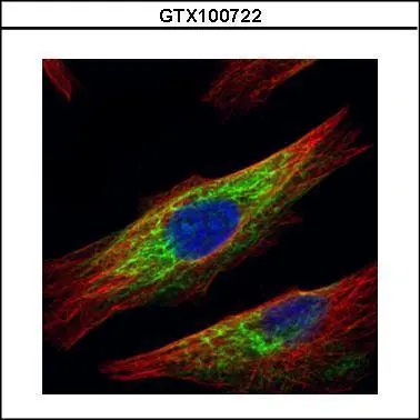
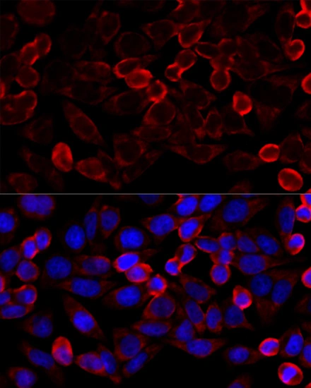
Confocal immunofluorescence analysis (Olympus FV10i) of paraformaldehyde-fixed HeLa, using HDAC6(GTX100722) antibody (Green) at 1:500 dilution. Alpha-tubulin filaments were labeled with GTX11304 (Red) at 1:2000.
 and HDAC6-transfected (+, including 3xFlag-tag) 293T whole cell extracts (30 μg) were separated by 5% SDS-PAGE, and the membrane was blotted with HDAC6 antibody (GTX100722) diluted by 1:5000.")

![Various whole cell extracts (30 μg) were separated by 5% SDS-PAGE, and the membrane was blotted with HDAC6 antibody [N1], N-term (GTX100722) diluted at 1:1000. The HRP-conjugated anti-rabbit IgG antibody (GTX213110-01) was used to detect the primary antibody. Corresponding RNA expression data for the same cell lines are based on Human Protein Atlas program.](https://www.genetex.com/upload/website/prouct_img/normal/GTX100722/GTX100722_40058_20250207_WB_TPM_watermark_25021320_453.webp "Various whole cell extracts (30 μg) were separated by 5% SDS-PAGE, and the membrane was blotted with HDAC6 antibody [N1], N-term (GTX100722) diluted at 1:1000. The HRP-conjugated anti-rabbit IgG antibody (GTX213110-01) was used to detect the primary antibody. Corresponding RNA expression data for the same cell lines are based on Human Protein Atlas program.")
Confocal immunofluorescence analysis (Olympus FV10i) of paraformaldehyde-fixed HeLa, using HDAC6(GTX100722) antibody (Green) at 1:500 dilution. Alpha-tubulin filaments were labeled with GTX11304 (Red) at 1:2000.
HDAC6 antibody [N1], N-term
GTX100722
ApplicationsFlow Cytometry, ImmunoFluorescence, Western Blot, ImmunoCytoChemistry, ImmunoHistoChemistry, ImmunoHistoChemistry Paraffin
Product group Antibodies
ReactivityHuman, Rat
TargetHDAC6
Overview
- SupplierGeneTex
- Product NameHDAC6 antibody [N1], N-term
- Delivery Days Customer9
- Application Supplier NoteWB: 1:1000-1:10000. ICC/IF: 1:100-1:1000. *Optimal dilutions/concentrations should be determined by the researcher.Not tested in other applications.
- ApplicationsFlow Cytometry, ImmunoFluorescence, Western Blot, ImmunoCytoChemistry, ImmunoHistoChemistry, ImmunoHistoChemistry Paraffin
- CertificationResearch Use Only
- ClonalityPolyclonal
- Concentration0.18 mg/ml
- ConjugateUnconjugated
- Gene ID10013
- Target nameHDAC6
- Target descriptionhistone deacetylase 6
- Target synonymsCPBHM, HD6, JM21, KDAC6, PPP1R90, protein deacetylase HDAC6, E3 ubiquitin-protein ligase HDAC6, alpha-tubulin deacetylase HDAC6, protein phosphatase 1, regulatory subunit 90, tubulin-lysine deacetylase HDAC6
- HostRabbit
- IsotypeIgG
- Protein IDQ9UBN7
- Protein NameProtein deacetylase HDAC6
- Scientific DescriptionHistones play a critical role in transcriptional regulation, cell cycle progression, and developmental events. Histone acetylation/deacetylation alters chromosome structure and affects transcription factor access to DNA. The protein encoded by this gene belongs to class II of the histone deacetylase/acuc/apha family. It contains an internal duplication of two catalytic domains which appear to function independently of each other. This protein possesses histone deacetylase activity and represses transcription. [provided by RefSeq]
- ReactivityHuman, Rat
- Storage Instruction-20°C or -80°C,2°C to 8°C
- UNSPSC41116161
Datasheet
Related products
Product group Antibodies
Anti-HDAC6 AntibodyA97491
ApplicationsWestern Blot, ELISA, ImmunoHistoChemistry
ReactivityHuman, Mouse
- SizePrice
Product group Antibodies
Anti-HDAC6 Antibody144-01732
ApplicationsWestern Blot, ImmunoHistoChemistry
ReactivityHuman, Mouse, Rat
TargetHDAC6
- SizePrice
Product group Antibodies

HDAC6 Antibody (Ser22)LS-C769272
ApplicationsImmunoFluorescence, Western Blot, ELISA, ImmunoHistoChemistry, ImmunoHistoChemistry Paraffin
ReactivityHuman, Mouse
TargetHDAC6
- SizePrice
Product group Antibodies
HDAC6 Polyclonal AntibodyBS-2811R
ApplicationsImmunoFluorescence, Western Blot, ELISA, ImmunoCytoChemistry, ImmunoHistoChemistry, ImmunoHistoChemistry Frozen, ImmunoHistoChemistry Paraffin
ReactivityBovine, Equine, Human, Mouse, Rabbit, Rat
TargetHDAC6
- SizePrice
Product group Antibodies
HDAC6 AntibodyCSB-PA002895
ApplicationsImmunoFluorescence, Western Blot, ELISA, ImmunoHistoChemistry
ReactivityHuman, Mouse
TargetHDAC6
- SizePrice
Product group Antibodies

Goat anti-HDAC6EB05431
ApplicationsFlow Cytometry, ImmunoFluorescence, ELISA, ImmunoHistoChemistry
ReactivityBovine, Canine, Human, Mouse, Rat
TargetHDAC6
- SizePrice
Product group Antibodies
ApplicationsFlow Cytometry
TargetHDAC6
- SizePrice
Product group Antibodies
HDAC6 antibodyGTX02885
ApplicationsImmunoFluorescence, ImmunoPrecipitation, Western Blot, ImmunoCytoChemistry, ImmunoHistoChemistry, ImmunoHistoChemistry Paraffin
ReactivityHuman, Mouse
TargetHDAC6
- SizePrice
![HDAC6 antibody [C3], C-term detects HDAC6 protein by western blot analysis. Non-transfected (-) and HDAC6-transfected (+) 293T whole cell extracts (30 μg) were separated by 5% SDS-PAGE, and the membrane was blotted with HDAC6 antibody [C3], C-term (GTX100677) diluted at 1:500.](https://www.genetex.com/upload/website/prouct_img/normal/GTX100677/GTX100677_40058_20150917_WB_B_w_23060100_123.webp)
Product group Antibodies
HDAC6 antibody [C3], C-termGTX100677
ApplicationsWestern Blot
ReactivityHuman
TargetHDAC6
- SizePrice
![WB analysis of HEK293T cells transfected with HDAC6 plasmid (Right) or empty vector (Left) for 48 hrs using GTX84377 HDAC6 antibody [4C5]. Loading : 5 ug per lane](https://www.genetex.com/upload/website/prouct_img/normal/GTX84377/GTX84377_4324_WB_w_23061420_795.webp)
Product group Antibodies
References
HDAC6 antibody [4C5]GTX84377
ApplicationsFlow Cytometry, ImmunoFluorescence, Western Blot, ImmunoCytoChemistry, ImmunoHistoChemistry, ImmunoHistoChemistry Paraffin
ReactivityCanine, Human, Monkey, Mouse, Rat
TargetHDAC6
- SizePrice