

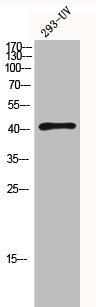
Western Blot analysis of 293-UV cells using HDAC8 Polyclonal Antibody

Western Blot analysis of 293-UV cells using HDAC8 Polyclonal Antibody
HDAC8 Antibody
CSB-PA002898
ApplicationsWestern Blot, ELISA, ImmunoHistoChemistry
Product group Antibodies
ReactivityHuman, Monkey, Mouse, Rat
TargetHDAC8
Overview
- SupplierCusabio
- Product NameHDAC8 Antibody
- Delivery Days Customer20
- ApplicationsWestern Blot, ELISA, ImmunoHistoChemistry
- CertificationResearch Use Only
- ClonalityPolyclonal
- ConjugateUnconjugated
- Gene ID55869
- Target nameHDAC8
- Target descriptionhistone deacetylase 8
- Target synonymsCDA07, CDLS5, HD8, HDACL1, KDAC8, MRXS6, RPD3, WTS, histone deacetylase 8, histone deacetylase-like 1, protein deacetylase HDAC8, protein decrotonylase HDAC8
- HostRabbit
- IsotypeIgG
- Protein IDQ9BY41
- Protein NameHistone deacetylase 8
- ReactivityHuman, Monkey, Mouse, Rat
- Storage Instruction-20°C or -80°C
- UNSPSC41116161
Related products
Product group Antibodies
Anti-HDAC8 AntibodyA95865
ApplicationsWestern Blot, ELISA, ImmunoHistoChemistry
ReactivityHuman, Mouse, Rat
- SizePrice
Product group Antibodies
Anti-HDAC8 Antibody144-05829
ApplicationsImmunoFluorescence, Western Blot, ImmunoHistoChemistry
ReactivityHuman, Mouse, Rat
TargetHDAC8
- SizePrice

Product group Antibodies
Anti-HDAC8 Antibody Picoband(r)A01843-1-CARRIER-FREE
ApplicationsWestern Blot, ELISA
ReactivityHuman, Mouse, Rat
TargetHDAC8
- SizePrice
Product group Antibodies
HDAC8 Recombinant AntibodyBSM-52088R
ApplicationsFlow Cytometry, ImmunoFluorescence, Western Blot, ImmunoCytoChemistry
ReactivityHuman, Mouse, Rat
TargetHDAC8
- SizePrice
Product group Antibodies
HDAC8 Polyclonal AntibodyCAC14844
ApplicationsWestern Blot, ELISA
ReactivityRat
TargetHDAC8
- SizePrice
Product group Antibodies

HDAC8 AntibodyLS-C400791
ApplicationsELISA, ImmunoHistoChemistry
ReactivityHuman, Mouse, Rat
TargetHDAC8
- SizePrice

Product group Antibodies
Anti-HDAC8 AntibodyHPA048560
ApplicationsImmunoCytoChemistry, ImmunoHistoChemistry
ReactivityHuman
TargetHDAC8
- SizePrice
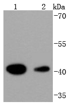
![Non-transfected (–) and transfected (+) 293T whole cell extracts (30 μg) were separated by 10% SDS-PAGE, and the membrane was blotted with HDAC8 antibody [N1C2] (GTX105074) diluted at 1:500. The HRP-conjugated anti-rabbit IgG antibody (GTX213110-01) was used to detect the primary antibody.](https://www.genetex.com/upload/website/prouct_img/normal/GTX105074/GTX105074_40023_20190614_WB_shRNA_watermark_w_23060120_640.webp)
Product group Antibodies
HDAC8 antibody [N1C2]GTX105074
ApplicationsWestern Blot
ReactivityHuman
TargetHDAC8
- SizePrice