

HDAC8 antibody [HDAC8-48]
GTX12176
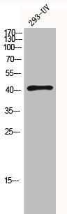
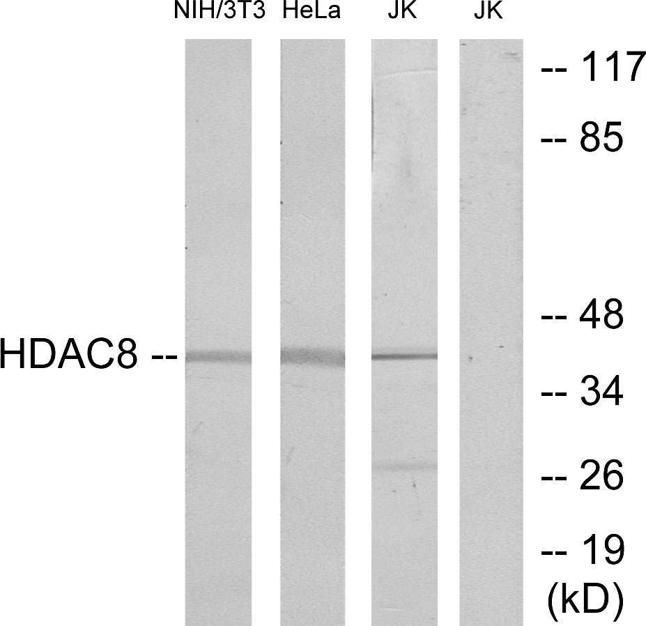
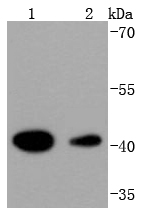
ApplicationsWestern Blot, ELISA
Product group Antibodies
ReactivityHuman
TargetHDAC8
Overview
- SupplierGeneTex
- Product NameHDAC8 antibody [HDAC8-48]
- Delivery Days Customer9
- Application Supplier NoteWB: 4 microg/ml. *Optimal dilutions/concentrations should be determined by the researcher.Not tested in other applications.
- ApplicationsWestern Blot, ELISA
- CertificationResearch Use Only
- ClonalityMonoclonal
- Clone IDHDAC8-48
- Concentration2.0-2.5 mg/ml
- ConjugateUnconjugated
- Gene ID55869
- Target nameHDAC8
- Target descriptionhistone deacetylase 8
- Target synonymsCDA07, CDLS5, HD8, HDACL1, KDAC8, MRXS6, RPD3, WTS, histone deacetylase 8, histone deacetylase-like 1, protein deacetylase HDAC8, protein decrotonylase HDAC8
- HostMouse
- IsotypeIgG1
- Protein IDQ9BY41
- Protein NameHistone deacetylase 8
- Scientific DescriptionRegulation of gene expression is mediated by several mechanisms such as DNA methylation, ATP-dependent chromatin remodeling, and post translational modifications of histones, which include the dynamic acetylation and deacetylation of epsilon-amino groups of lysine residues present in the tail of core histones. The enzymes responsible for reversible acetylation/deacetylation processes are histone acetyltransferases(HATs) and histone deacetylases (HDACs), respectively. HATs act as transcriptional coactivators, and HDACs are part of transcriptional corepressor complexes. Mammalian HDACs can be divided into three classes according to sequence homology. Class I consists of the yeast Rpd3-like proteins (HDAC1, HDAC2, HDAC3, and HDAC8). Class II consists of the yeast Hda1-like proteins (HDAC4, HDAC5, HDAC6, HDAC7, HDAC9, and HDAC10). Class III comprises the yeast Sir2-like proteins. Class I HDACs are ubiquitously expressed, and most class II HDACs are tissue-specific. Class II HDACs have been implicated in the regulation of muscle differentiation. Interaction of HDAC4, 5, and 7 with members of the MEF2 family of transcription factors represses their transcriptional activity and prevents myogenesis. The deacetylase activity of class II HDACs is regulated by subcellular localization. The HDAC8 gene encodes for a 377 amino acid protein with a molecular weight of 43 kDa and is localized within the nucleus. HDAC8 mRNA is expressed in heart, lung, kidney, and pancreas as well as in several cell lines derived from cancer tissues.
- ReactivityHuman
- Storage Instruction-20°C or -80°C,2°C to 8°C
- UNSPSC12352203
Datasheet
Related products
Product group Antibodies
HDAC8 AntibodyCSB-PA002898
ApplicationsWestern Blot, ELISA, ImmunoHistoChemistry
ReactivityHuman, Monkey, Mouse, Rat
TargetHDAC8
- SizePrice
Product group Antibodies
HDAC8 Polyclonal AntibodyCAC14844
ApplicationsWestern Blot, ELISA
ReactivityRat
TargetHDAC8
- SizePrice

Product group Antibodies
Anti-HDAC8 Antibody Picoband(r)A01843-1-CARRIER-FREE
ApplicationsWestern Blot, ELISA
ReactivityHuman, Mouse, Rat
TargetHDAC8
- SizePrice
Product group Antibodies
Anti-HDAC8 Antibody144-05829
ApplicationsImmunoFluorescence, Western Blot, ImmunoHistoChemistry
ReactivityHuman, Mouse, Rat
TargetHDAC8
- SizePrice
Product group Antibodies
Anti-HDAC8 AntibodyA95865
ApplicationsWestern Blot, ELISA, ImmunoHistoChemistry
ReactivityHuman, Mouse, Rat
- SizePrice

Product group Antibodies
Anti-HDAC8 AntibodyHPA048560
ApplicationsImmunoCytoChemistry, ImmunoHistoChemistry
ReactivityHuman
TargetHDAC8
- SizePrice
Product group Antibodies

HDAC8 AntibodyLS-C400791
ApplicationsELISA, ImmunoHistoChemistry
ReactivityHuman, Mouse, Rat
TargetHDAC8
- SizePrice
Product group Antibodies
HDAC8 Recombinant AntibodyBSM-52088R
ApplicationsFlow Cytometry, ImmunoFluorescence, Western Blot, ImmunoCytoChemistry
ReactivityHuman, Mouse, Rat
TargetHDAC8
- SizePrice
![Non-transfected (–) and transfected (+) 293T whole cell extracts (30 μg) were separated by 10% SDS-PAGE, and the membrane was blotted with HDAC8 antibody [N1C2] (GTX105074) diluted at 1:500. The HRP-conjugated anti-rabbit IgG antibody (GTX213110-01) was used to detect the primary antibody.](https://www.genetex.com/upload/website/prouct_img/normal/GTX105074/GTX105074_40023_20190614_WB_shRNA_watermark_w_23060120_640.webp)
Product group Antibodies
HDAC8 antibody [N1C2]GTX105074
ApplicationsWestern Blot
ReactivityHuman
TargetHDAC8
- SizePrice