

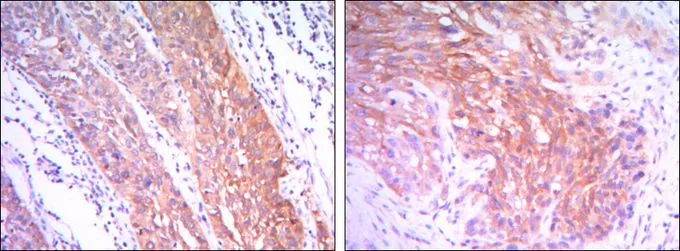
IHC-P analysis of esophagus cancer tissue (left) and human lung cancer (right) using GTX82791 Hexokinase II antibody [3D3].
![WB analysis of Jurkat (1), HeLa (2) and HEK293 (3) cell lysate using GTX82791 Hexokinase II antibody [3D3].](https://www.genetex.com/upload/website/prouct_img/normal/GTX82791/GTX82791_20170912_WB_w_23051501_734.webp "WB analysis of Jurkat (1), HeLa (2) and HEK293 (3) cell lysate using GTX82791 Hexokinase II antibody [3D3].")
![FACS analysis of K562 cells using GTX82791 Hexokinase II antibody [3D3]. Green : Hexokinase II Purple : negative control](https://www.genetex.com/upload/website/prouct_img/normal/GTX82791/GTX82791_20170912_FACS_w_23051501_847.webp "FACS analysis of K562 cells using GTX82791 Hexokinase II antibody [3D3]. Green : Hexokinase II Purple : negative control")
IHC-P analysis of esophagus cancer tissue (left) and human lung cancer (right) using GTX82791 Hexokinase II antibody [3D3].
Hexokinase II antibody [3D3]
GTX82791
ApplicationsFlow Cytometry, Western Blot, ELISA, ImmunoHistoChemistry, ImmunoHistoChemistry Paraffin
Product group Antibodies
ReactivityHuman
TargetHK2
Overview
- SupplierGeneTex
- Product NameHexokinase II antibody [3D3]
- Delivery Days Customer9
- Application Supplier NoteWB: 1/500 - 1/2000. IHC-P: 1/200 - 1/1000. FCM: 1/200 - 1/400. ELISA: 1/10000. *Optimal dilutions/concentrations should be determined by the researcher.Not tested in other applications.
- ApplicationsFlow Cytometry, Western Blot, ELISA, ImmunoHistoChemistry, ImmunoHistoChemistry Paraffin
- CertificationResearch Use Only
- ClonalityMonoclonal
- Clone ID3D3
- ConjugateUnconjugated
- Gene ID3099
- Target nameHK2
- Target descriptionhexokinase 2
- Target synonymsHKII, HXK2, hexokinase-2, hexokinase type II, hexokinase-2, muscle, hexokinase-B, muscle form hexokinase
- HostMouse
- IsotypeIgG1
- Protein IDP52789
- Protein NameHexokinase-2
- Scientific DescriptionHexokinases phosphorylate glucose to produce glucose-6-phosphate, the first step in most glucose metabolism pathways. This gene encodes hexokinase 2, the predominant form found in skeletal muscle. It localizes to the outer membrane of mitochondria. Expression of this gene is insulin-responsive, and studies in rat suggest that it is involved in the increased rate of glycolysis seen in rapidly growing cancer cells. [provided by RefSeq, Apr 2009]
- ReactivityHuman
- Storage Instruction-20°C or -80°C,2°C to 8°C
- UNSPSC41116161
Datasheet
Related products
Product group Antibodies
ApplicationsImmunoFluorescence, Western Blot, ImmunoCytoChemistry, ImmunoHistoChemistry
ReactivityHuman, Mouse, Rat
- SizePrice

Product group Antibodies
Anti-Hexokinase II/HK2 Antibody Picoband(r)A01389-CARRIER-FREE
ApplicationsFlow Cytometry, Western Blot, ImmunoHistoChemistry
ReactivityHuman, Mouse, Rat
TargetHK2
- SizePrice
Product group Antibodies
Anti-HK2 Antibody144-00994
ApplicationsImmunoFluorescence, Western Blot, ImmunoHistoChemistry
ReactivityHuman, Mouse, Rat
TargetHK2
- SizePrice
Product group Antibodies
References
ApplicationsImmunoFluorescence, Western Blot, ImmunoCytoChemistry, ImmunoHistoChemistry, ImmunoHistoChemistry Paraffin
ReactivityBovine, Canine, Human, Mouse, Porcine, Rabbit, Rat, Sheep
TargetHK2
- SizePrice

Product group Antibodies
HK2 AntibodyCSB-PA010469LA01HU
ApplicationsImmunoFluorescence, Western Blot, ELISA
ReactivityHuman
TargetHK2
- SizePrice
Product group Antibodies
HK2 Polyclonal AntibodyCAC14936
ApplicationsImmunoFluorescence, Western Blot, ELISA
TargetHK2
- SizePrice
Product group Antibodies

HK2 / Hexokinase 2 AntibodyLS-C401376
ApplicationsELISA, ImmunoHistoChemistry
ReactivityHuman, Mouse, Rat
TargetHK2
- SizePrice
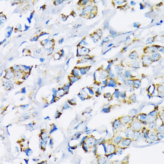
![ICC/IF analysis of HepG2 cells using GTX84357 Hexokinase II antibody [4C5].](https://www.genetex.com/upload/website/prouct_img/normal/GTX84357/GTX84357_1055_ICCIF_w_23061420_212.webp)
Product group Antibodies
Hexokinase II antibody [4C5]GTX84357
ApplicationsFlow Cytometry, ImmunoFluorescence, Western Blot, ImmunoCytoChemistry
ReactivityCanine, Human
TargetHK2
- SizePrice

Product group Antibodies
Anti-HK2 AntibodyHPA028587
ApplicationsWestern Blot
ReactivityHuman
TargetHK2
- SizePrice
Product group Antibodies
Hexokinase II antibodyGTX111525
ApplicationsImmunoFluorescence, Western Blot, ImmunoCytoChemistry, ImmunoHistoChemistry, ImmunoHistoChemistry Paraffin
ReactivityHuman, Mouse, Rat
TargetHK2
- SizePrice