

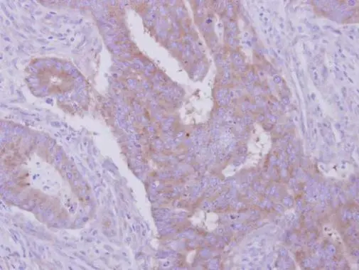
Immunohistochemical analysis of paraffin-embedded human colon carcinoma, using HGS(GTX101718) antibody at 1:500 dilution.
Antigen Retrieval: Trilogy? (EDTA based, pH 8.0) buffer, 15min
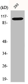
![Various whole cell extracts (30 μg) were separated by 7.5% SDS-PAGE, and the membrane was blotted with HGS antibody [C2C3], C-term (GTX101718) diluted at 1:1000. The HRP-conjugated anti-rabbit IgG antibody (GTX213110-01) was used to detect the primary antibody.](https://www.genetex.com/upload/website/prouct_img/normal/GTX101718/GTX101718_39876_20210716_WB_w_23060100_815.webp "Various whole cell extracts (30 μg) were separated by 7.5% SDS-PAGE, and the membrane was blotted with HGS antibody [C2C3], C-term (GTX101718) diluted at 1:1000. The HRP-conjugated anti-rabbit IgG antibody (GTX213110-01) was used to detect the primary antibody.")
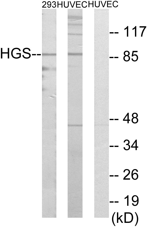
![Various tissue extracts (50 μg) were separated by 7.5% SDS-PAGE, and the membrane was blotted with HGS antibody [C2C3], C-term (GTX101718) diluted at 1:1000. The HRP-conjugated anti-rabbit IgG antibody (GTX213110-01) was used to detect the primary antibody.](https://www.genetex.com/upload/website/prouct_img/normal/GTX101718/GTX101718_39876_20220311_WB_M_R_w_23060100_792.webp "Various tissue extracts (50 μg) were separated by 7.5% SDS-PAGE, and the membrane was blotted with HGS antibody [C2C3], C-term (GTX101718) diluted at 1:1000. The HRP-conjugated anti-rabbit IgG antibody (GTX213110-01) was used to detect the primary antibody.")
 of methanol-fixed A431, using HGS(GTX101718) antibody (green) at 1:200 dilution. Alpha-tubulin filaments were labeled with GTX11304 (red) at 1:500.")

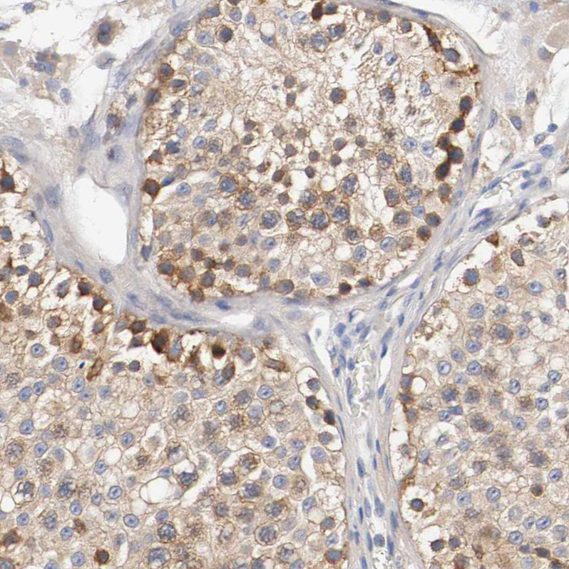
Immunohistochemical analysis of paraffin-embedded human colon carcinoma, using HGS(GTX101718) antibody at 1:500 dilution.
Antigen Retrieval: Trilogy? (EDTA based, pH 8.0) buffer, 15min
HGS antibody [C2C3], C-term
GTX101718
ApplicationsImmunoFluorescence, Western Blot, ImmunoCytoChemistry, ImmunoHistoChemistry, ImmunoHistoChemistry Paraffin
Product group Antibodies
ReactivityHuman, Mouse, Rat
TargetHGS
Overview
- SupplierGeneTex
- Product NameHGS antibody [C2C3], C-term
- Delivery Days Customer9
- Application Supplier NoteWB: 1:1000-1:10000. ICC/IF: 1:100-1:1000. IHC-P: 1:100-1:1000. *Optimal dilutions/concentrations should be determined by the researcher.Not tested in other applications.
- ApplicationsImmunoFluorescence, Western Blot, ImmunoCytoChemistry, ImmunoHistoChemistry, ImmunoHistoChemistry Paraffin
- CertificationResearch Use Only
- ClonalityPolyclonal
- Concentration1 mg/ml
- ConjugateUnconjugated
- Gene ID9146
- Target nameHGS
- Target descriptionhepatocyte growth factor-regulated tyrosine kinase substrate
- Target synonymsHRS, hepatocyte growth factor-regulated tyrosine kinase substrate, human growth factor-regulated tyrosine kinase substrate, protein pp110
- HostRabbit
- IsotypeIgG
- Protein IDO14964
- Protein NameHepatocyte growth factor-regulated tyrosine kinase substrate
- Scientific DescriptionThe protein encoded by this gene regulates endosomal sorting and plays a critical role in the recycling and degradation of membrane receptors. The encoded protein sorts monoubiquitinated membrane proteins into the multivesicular body, targeting these proteins for lysosome-dependent degradation. [provided by RefSeq]
- ReactivityHuman, Mouse, Rat
- Storage Instruction-20°C or -80°C,2°C to 8°C
- UNSPSC41116161
Datasheet
Related products
Product group Antibodies
HGS AntibodyCSB-PA002970
ApplicationsImmunoFluorescence, Western Blot, ELISA, ImmunoHistoChemistry
ReactivityHuman, Mouse, Rat
TargetHGS
- SizePrice
Product group Antibodies
Anti-HGS AntibodyA95484
ApplicationsImmunoFluorescence, Western Blot, ELISA, ImmunoHistoChemistry
ReactivityHuman, Mouse, Rat
- SizePrice

Product group Antibodies
Anti-HGS Antibody Picoband(r)A01174-1-CARRIER-FREE
ApplicationsFlow Cytometry, ImmunoFluorescence, Western Blot, ELISA, ImmunoCytoChemistry, ImmunoHistoChemistry
ReactivityHuman, Mouse, Rat
TargetHGS
- SizePrice
Product group Antibodies

HRS AntibodyABX430007
ApplicationsWestern Blot, ELISA, ImmunoHistoChemistry
- SizePrice
Product group Antibodies

Goat anti-HRS / HGSEB07211
ApplicationsWestern Blot, ELISA, ImmunoHistoChemistry
ReactivityCanine, Human, Mouse, Rat
TargetHGS
- SizePrice
Product group Antibodies
Anti-HGS AntibodyHPA004872
ApplicationsWestern Blot, ImmunoCytoChemistry, ImmunoHistoChemistry
ReactivityHuman
TargetHGS
- SizePrice
Product group Antibodies

HRS / HGS AntibodyLS-C408351
ApplicationsWestern Blot
ReactivityHuman, Mouse, Rat
TargetHGS
- SizePrice
Product group Antibodies
ApplicationsImmunoPrecipitation, Western Blot, ImmunoCytoChemistry, ImmunoHistoChemistry
TargetHGS
- SizePrice
![Whole cell extract (30 μg) was separated by 7.5% SDS-PAGE, and the membrane was blotted with HGS antibody [HL1730] (GTX637378) diluted at 1:1000. The HRP-conjugated anti-rabbit IgG antibody (GTX213110-01) was used to detect the primary antibody.](https://www.genetex.com/upload/website/prouct_img/normal/GTX637378/GTX637378_44830_20221014_WB_22101718_254.webp)
Product group Antibodies
HGS antibody [HL1730]GTX637378
ApplicationsImmunoFluorescence, Western Blot, ImmunoCytoChemistry, ImmunoHistoChemistry, ImmunoHistoChemistry Paraffin
ReactivityCanine, Feline, Human
TargetHGS
- SizePrice