

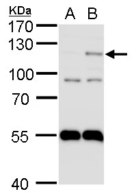
WB analysis of hypoxia-treated A549 cell lysate using GTX30123 HIF2 alpha antibody [ep190b]. Lane 1: normoxic A549 lysate control Lane 2: hypoxic A549 lysate
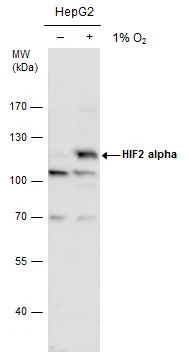
![WB analysis of multiple samples using GTX30123 HIF2 alpha antibody [ep190b]. Lane 1 and 5: HepG2 without Cobalt (II) Chloride Lane 2 and 6: HepG2 with Cobalt (II) Chloride Lane 3 and 7: HepG2 normoxic Lane 4 and 8 : HepG2 hypoxic Dilution : 1-2 μg/ml](https://www.genetex.com/upload/website/prouct_img/normal/GTX30123/GTX30123_1337_WB_w_23060722_196.webp "WB analysis of multiple samples using GTX30123 HIF2 alpha antibody [ep190b]. Lane 1 and 5: HepG2 without Cobalt (II) Chloride Lane 2 and 6: HepG2 with Cobalt (II) Chloride Lane 3 and 7: HepG2 normoxic Lane 4 and 8 : HepG2 hypoxic Dilution : 1-2 μg/ml")
![FACS analysis of HepG2 cells using GTX30123 HIF2 alpha antibody [ep190b]. M1 is defined by unstained cells. Purple : primary antibody Dilution : 1:400](https://www.genetex.com/upload/website/prouct_img/normal/GTX30123/GTX30123_28_FACS_w_23060722_106.webp "FACS analysis of HepG2 cells using GTX30123 HIF2 alpha antibody [ep190b]. M1 is defined by unstained cells. Purple : primary antibody Dilution : 1:400")
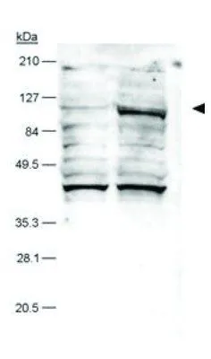
![WB analysis of HeLa cell lysate using GTX30123 HIF2 alpha antibody [ep190b].](https://www.genetex.com/upload/website/prouct_img/normal/GTX30123/GTX30123_1336_WB_w_23060722_693.webp "WB analysis of HeLa cell lysate using GTX30123 HIF2 alpha antibody [ep190b].")
![IHC-P analysis of human heart tissue using GTX30123 HIF2 alpha antibody [ep190b].](https://www.genetex.com/upload/website/prouct_img/normal/GTX30123/GTX30123_1609_IHC_w_23060722_954.webp "IHC-P analysis of human heart tissue using GTX30123 HIF2 alpha antibody [ep190b].")


WB analysis of hypoxia-treated A549 cell lysate using GTX30123 HIF2 alpha antibody [ep190b]. Lane 1: normoxic A549 lysate control Lane 2: hypoxic A549 lysate
HIF2 alpha antibody [ep190b]
GTX30123
ApplicationsGel Shift Assay, Flow Cytometry, ImmunoFluorescence, ImmunoPrecipitation, Western Blot, ChIP Chromatin ImmunoPrecipitation, ELISA, ImmunoCytoChemistry, ImmunoHistoChemistry, ImmunoHistoChemistry Paraffin
Product group Antibodies
ReactivityHamster, Human, Mouse, Rat
TargetEPAS1
Overview
- SupplierGeneTex
- Product NameHIF2 alpha antibody [ep190b]
- Delivery Days Customer9
- Application Supplier NoteWB: 1 - 2 microg/ml. IHC-P: 1:150 - 1:300. FCM: 1:400. ELISA: 1:100 - 1:2000. IHC: 1:150 - 1:300. *Optimal dilutions/concentrations should be determined by the researcher.Not tested in other applications.
- ApplicationsGel Shift Assay, Flow Cytometry, ImmunoFluorescence, ImmunoPrecipitation, Western Blot, ChIP Chromatin ImmunoPrecipitation, ELISA, ImmunoCytoChemistry, ImmunoHistoChemistry, ImmunoHistoChemistry Paraffin
- CertificationResearch Use Only
- ClonalityMonoclonal
- Clone IDep190b
- Concentration1 mg/ml
- ConjugateUnconjugated
- Gene ID2034
- Target nameEPAS1
- Target descriptionendothelial PAS domain protein 1
- Target synonymsECYT4, HIF2A, HLF, MOP2, PASD2, bHLHe73, endothelial PAS domain-containing protein 1, EPAS-1, HIF-1-alpha-like factor, HIF-1alpha-like factor, HIF-2-alpha, HIF2-alpha, PAS domain-containing protein 2, basic-helix-loop-helix-PAS protein MOP2, class E basic helix-loop-helix protein 73, hypoxia-inducible factor 2 alpha, member of PAS protein 2
- HostMouse
- IsotypeIgG1
- Protein IDQ99814
- Protein NameEndothelial PAS domain-containing protein 1
- Scientific DescriptionThis gene encodes a transcription factor involved in the induction of genes regulated by oxygen, which is induced as oxygen levels fall. The encoded protein contains a basic-helix-loop-helix domain protein dimerization domain as well as a domain found in proteins in signal transduction pathways which respond to oxygen levels. Mutations in this gene are associated with erythrocytosis familial type 4. [provided by RefSeq]
- ReactivityHamster, Human, Mouse, Rat
- Storage Instruction-20°C or -80°C,2°C to 8°C
- UNSPSC41116161
References
- Changes in cell fate determine the regenerative and functional capacity of the developing kidney before and after release of obstruction. Nagalakshmi VK et al., 2018 Dec 12, Clin Sci (Lond)Read this paper
- Thiamine deficiency activates hypoxia inducible factor-1alpha to facilitate pro-apoptotic responses in mouse primary astrocytes. Zera K et al., 2017, PLoS OneRead this paper
- VHL and PTEN loss coordinate to promote mouse liver vascular lesions. Chen S et al., 2010 Mar, AngiogenesisRead this paper
- Identification of Ror2 as a hypoxia-inducible factor target in von Hippel-Lindau-associated renal cell carcinoma. Wright TM et al., 2010 Apr 23, J Biol ChemRead this paper
- ATR and Chk1 suppress a caspase-3-dependent apoptotic response following DNA replication stress. Myers K et al., 2009 Jan, PLoS GenetRead this paper
- VHL type 2B mutations retain VBC complex form and function. Hacker KE et al., 2008, PLoS OneRead this paper
- Hypoxia-inducible factor-1alpha and hypoxia-inducible factor-2alpha are expressed in kaposi sarcoma and modulated by insulin-like growth factor-I. Catrina SB et al., 2006 Aug 1, Clin Cancer ResRead this paper
Datasheet
Related products
Product group Antibodies
EPAS1 AntibodyCSB-PA005478
ApplicationsWestern Blot, ELISA
ReactivityHuman, Mouse, Rat
TargetEPAS1
- SizePrice

Product group Antibodies
Anti-HIF-2-alpha/EPAS1 Antibody Picoband(r)A01248-1-CARRIER-FREE
ApplicationsFlow Cytometry, Western Blot
ReactivityHuman, Mouse, Rat
TargetEPAS1
- SizePrice

Product group Antibodies
Anti-EPAS1 AntibodyHPA031200
ApplicationsImmunoCytoChemistry
ReactivityHuman
TargetEPAS1
- SizePrice
Product group Antibodies

HIF2A / EPAS1 AntibodyLS-C409163
ApplicationsImmunoFluorescence, Western Blot
ReactivityHuman
TargetEPAS1
- SizePrice
Product group Antibodies
ApplicationsImmunoPrecipitation, Western Blot, ImmunoCytoChemistry, ImmunoHistoChemistry
ReactivityMouse, Porcine
TargetEPAS1
- SizePrice
Product group Antibodies
HIF-2 alpha Recombinant AntibodyBSM-54278R
ApplicationsFlow Cytometry, ImmunoFluorescence, Western Blot, ImmunoCytoChemistry
ReactivityHuman, Mouse, Rat
TargetEPAS1
- SizePrice
Product group Antibodies
HIF2 alpha antibody [N1N3]GTX103707
ApplicationsWestern Blot, ChIP Chromatin ImmunoPrecipitation
ReactivityHuman, Zebra Fish
TargetEPAS1
- SizePrice
Product group Antibodies
HIF2 alpha antibodyGTX128793
ApplicationsImmunoPrecipitation, Western Blot
ReactivityHuman
TargetEPAS1
- SizePrice