

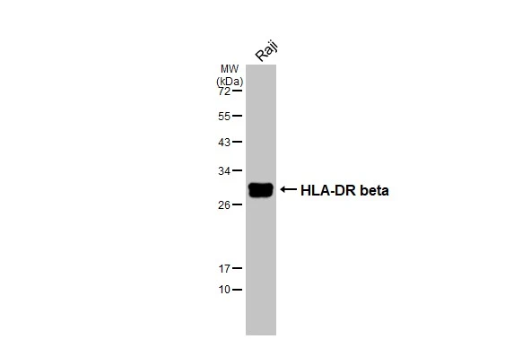
Whole cell extract (30 μg) was separated by 12% SDS-PAGE, and the membrane was blotted with HLA-DR beta antibody [N1C3] (GTX102047) diluted at 1:1000. The HRP-conjugated anti-rabbit IgG antibody (GTX213110-01) was used to detect the primary antibody.
Whole cell extract (30 μg) was separated by 12% SDS-PAGE, and the membrane was blotted with HLA-DR beta antibody [N1C3] (GTX102047) diluted at 1:1000. The HRP-conjugated anti-rabbit IgG antibody (GTX213110-01) was used to detect the primary antibody.
HLA-DR beta antibody [N1C3]
GTX102047
ApplicationsWestern Blot
Product group Antibodies
ReactivityHuman
TargetHLA-DRB1
Overview
- SupplierGeneTex
- Product NameHLA-DR beta antibody [N1C3]
- Delivery Days Customer9
- Application Supplier NoteWB: 1:500-1:3000. *Optimal dilutions/concentrations should be determined by the researcher.Not tested in other applications.
- ApplicationsWestern Blot
- CertificationResearch Use Only
- ClonalityPolyclonal
- Concentration1 mg/ml
- ConjugateUnconjugated
- Gene ID3123
- Target nameHLA-DRB1
- Target descriptionmajor histocompatibility complex, class II, DR beta 1
- Target synonymsDRB1, HLA-DR1B, HLA-DRB, SS1, major histocompatibility complex, class II, DR beta 1 precursor, HLA class II histocompatibility antigen, DR-1 beta chain, MHC class II HLA-DR beta 1 chain, human leucocyte antigen DRB1, lymphocyte antigen DRB1
- HostRabbit
- IsotypeIgG
- Protein IDP01911
- Protein NameHLA class II histocompatibility antigen, DRB1 beta chain
- ReactivityHuman
- Storage Instruction-20°C or -80°C,2°C to 8°C
- UNSPSC41116161
Datasheet
Related products
Product group Antibodies
Anti-HLA-DRB1 Antibody144-07685
ApplicationsImmunoFluorescence, Western Blot
ReactivityHuman, Mouse
TargetHLA-DRB1
- SizePrice
Product group Antibodies

HLA-DRB1 AntibodyLS-C830148
ApplicationsELISA, ImmunoHistoChemistry
ReactivityHuman
TargetHLA-DRB1
- SizePrice

Product group Antibodies
Anti-HLA Class II DRB1/HLA-DRB1 Antibody Picoband(r)A00568-CARRIER-FREE
ApplicationsWestern Blot
ReactivityHuman
TargetHLA-DRB1
- SizePrice
Product group Antibodies
HLA-DRB1 Polyclonal AntibodyCAC12865
ApplicationsImmunoFluorescence, Western Blot, ELISA
TargetHLA-DRB1
- SizePrice
Product group Antibodies
HLA-DRB1 AntibodyCSB-PA17949A0RB
ApplicationsELISA
ReactivityHuman
TargetHLA-DRB1
- SizePrice
![IHC-P analysis of human lung tissue using GTX04457 HLA-DRB1 antibody [MSVA-478R] HistoMAX?. Strong HLA-DRB1 staining in alveolar macrophages HLA-DRB1 immunohistochemistry.](https://www.genetex.com/upload/website/prouct_img/normal/GTX04457/GTX04457_20230728_IHC-P_63_23072722_466.webp)
Product group Antibodies
ApplicationsImmunoHistoChemistry, ImmunoHistoChemistry Paraffin
ReactivityHuman
TargetHLA-DRB1
- SizePrice
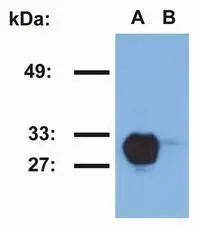
![WB analysis of Raji (A) and Jurkat (B) cell lysates using GTX78393 HLA-DR1 antibody [MEM-267].](https://www.genetex.com/upload/website/prouct_img/normal/GTX78393/GTX78393_20191028_WB_1_w_23061322_693.webp)
Product group Antibodies
HLA-DR1 antibody [MEM-267]GTX78393
ApplicationsFlow Cytometry, Western Blot, ELISA
ReactivityHuman
TargetHLA-DRB1
- SizePrice
Product group Antibodies
HLA-DR1 antibody [MEM-267] (PE)GTX79956
ApplicationsFlow Cytometry, Western Blot
ReactivityHuman
TargetHLA-DRB1
- SizePrice

Product group Antibodies
Anti-HLA-DRB1 AntibodyHPA043151
ApplicationsWestern Blot, ImmunoHistoChemistry
ReactivityHuman
TargetHLA-DRB1
- SizePrice
![HLA-DRB1 antibody [N1C3] detects HLA-DRB1 protein at cell membrane by immunofluorescent analysis. Sample: Raji cells were fixed in 4% paraformaldehyde at RT for 15 min. Green: HLA-DRB1 stained by HLA-DRB1 antibody [N1C3] (GTX104919) diluted at 1:500. Blue: Fluoroshield with DAPI (GTX30920).](https://www.genetex.com/upload/website/prouct_img/normal/GTX104919/GTX104919_43663_20220916_ICC_IF_22102723_790.webp)
Product group Antibodies
HLA-DRB1 antibody [N1C3]GTX104919
ApplicationsImmunoFluorescence, Western Blot, ImmunoCytoChemistry, ImmunoHistoChemistry, ImmunoHistoChemistry Paraffin
ReactivityHuman
TargetHLA-DRB1
- SizePrice