

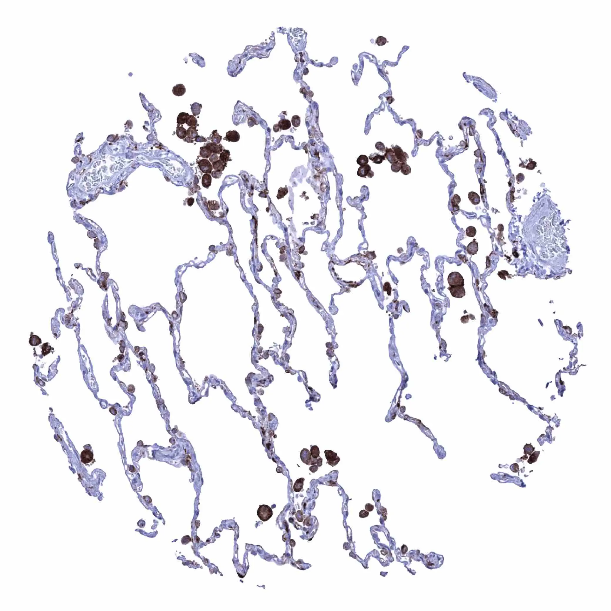
IHC-P analysis of human lung tissue using GTX04457 HLA-DRB1 antibody [MSVA-478R] HistoMAX?. Strong HLA-DRB1 staining in alveolar macrophages HLA-DRB1 immunohistochemistry.
![IHC-P analysis of human testicular seminoma tissue using GTX04457 HLA-DRB1 antibody [MSVA-478R] HistoMAX?. HLA-DRB1 negative seminoma showing HLA-DRB1 staining of inflammatory cells and of small vessels HLA-DRB1 immunohistochemistry.](https://www.genetex.com/upload/website/prouct_img/normal/GTX04457/GTX04457_20230728_IHC-P_193_23072723_202.webp "IHC-P analysis of human testicular seminoma tissue using GTX04457 HLA-DRB1 antibody [MSVA-478R] HistoMAX?. HLA-DRB1 negative seminoma showing HLA-DRB1 staining of inflammatory cells and of small vessels HLA-DRB1 immunohistochemistry.")
![IHC-P analysis of human liver tissue using GTX04457 HLA-DRB1 antibody [MSVA-478R] HistoMAX?. Intense HLA-DRB1 staining of inflammatory cells and of Kupffer cells HLA-DRB1 immunohistochemistry.](https://www.genetex.com/upload/website/prouct_img/normal/GTX04457/GTX04457_20230728_IHC-P_323_23072723_552.webp "IHC-P analysis of human liver tissue using GTX04457 HLA-DRB1 antibody [MSVA-478R] HistoMAX?. Intense HLA-DRB1 staining of inflammatory cells and of Kupffer cells HLA-DRB1 immunohistochemistry.")
IHC-P analysis of human lung tissue using GTX04457 HLA-DRB1 antibody [MSVA-478R] HistoMAX?. Strong HLA-DRB1 staining in alveolar macrophages HLA-DRB1 immunohistochemistry.
HLA-DRB1 antibody [MSVA-478R] HistoMAX(tm)
GTX04457
ApplicationsImmunoHistoChemistry, ImmunoHistoChemistry Paraffin
Product group Antibodies
ReactivityHuman
TargetHLA-DRB1
Overview
- SupplierGeneTex
- Product NameHLA-DRB1 antibody [MSVA-478R] HistoMAX(tm)
- Delivery Days Customer9
- Application Supplier NoteIHC-P: 1:100-1:200. *Optimal dilutions/concentrations should be determined by the researcher.Not tested in other applications.
- ApplicationsImmunoHistoChemistry, ImmunoHistoChemistry Paraffin
- CertificationResearch Use Only
- ClonalityMonoclonal
- Clone IDMSVA-478R
- Concentration0.2 mg/ml
- ConjugateUnconjugated
- Gene ID3123
- Target nameHLA-DRB1
- Target descriptionmajor histocompatibility complex, class II, DR beta 1
- Target synonymsDRB1, HLA-DR1B, HLA-DRB, SS1, major histocompatibility complex, class II, DR beta 1 precursor, HLA class II histocompatibility antigen, DR-1 beta chain, MHC class II HLA-DR beta 1 chain, human leucocyte antigen DRB1, lymphocyte antigen DRB1
- HostRabbit
- IsotypeIgG
- Protein IDP01911
- Protein NameHLA class II histocompatibility antigen, DRB1 beta chain
- Scientific DescriptionHLA-DRB1 belongs to the HLA class II beta chain paralogs. The class II molecule is a heterodimer consisting of an alpha (DRA) and a beta chain (DRB), both anchored in the membrane. It plays a central role in the immune system by presenting peptides derived from extracellular proteins. Class II molecules are expressed in antigen presenting cells (APC: B lymphocytes, dendritic cells, macrophages). The beta chain is approximately 26-28 kDa. It is encoded by 6 exons. Exon one encodes the leader peptide; exons 2 and 3 encode the two extracellular domains; exon 4 encodes the transmembrane domain; and exon 5 encodes the cytoplasmic tail. Within the DR molecule the beta chain contains all the polymorphisms specifying the peptide binding specificities. Hundreds of DRB1 alleles have been described and typing for these polymorphisms is routinely done for bone marrow and kidney transplantation. DRB1 is expressed at a level five times higher than its paralogs DRB3, DRB4 and DRB5. DRB1 is present in all individuals. Allelic variants of DRB1 are linked with either none or one of the genes DRB3, DRB4 and DRB5. There are 4 related pseudogenes: DRB2, DRB6, DRB7, DRB8 and DRB9. [provided by RefSeq, Jul 2008]
- ReactivityHuman
- Storage Instruction-20°C or -80°C,2°C to 8°C
- UNSPSC41116161
Datasheet
Related products
Product group Antibodies
Anti-HLA-DRB1 Antibody144-07685
ApplicationsImmunoFluorescence, Western Blot
ReactivityHuman, Mouse
TargetHLA-DRB1
- SizePrice
Product group Antibodies

HLA-DRB1 AntibodyLS-C830148
ApplicationsELISA, ImmunoHistoChemistry
ReactivityHuman
TargetHLA-DRB1
- SizePrice

Product group Antibodies
Anti-HLA Class II DRB1/HLA-DRB1 Antibody Picoband(r)A00568-CARRIER-FREE
ApplicationsWestern Blot
ReactivityHuman
TargetHLA-DRB1
- SizePrice
Product group Antibodies
HLA-DRB1 Polyclonal AntibodyCAC12865
ApplicationsImmunoFluorescence, Western Blot, ELISA
TargetHLA-DRB1
- SizePrice
Product group Antibodies
HLA-DRB1 AntibodyCSB-PA17949A0RB
ApplicationsELISA
ReactivityHuman
TargetHLA-DRB1
- SizePrice
![Whole cell extract (30 μg) was separated by 12% SDS-PAGE, and the membrane was blotted with HLA-DR beta antibody [N1C3] (GTX102047) diluted at 1:1000. The HRP-conjugated anti-rabbit IgG antibody (GTX213110-01) was used to detect the primary antibody.](https://www.genetex.com/upload/website/prouct_img/normal/GTX102047/GTX102047_39995_20240607_WB_24061301_543.webp)
Product group Antibodies
HLA-DR beta antibody [N1C3]GTX102047
ApplicationsWestern Blot
ReactivityHuman
TargetHLA-DRB1
- SizePrice
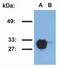
![WB analysis of Raji (A) and Jurkat (B) cell lysates using GTX78393 HLA-DR1 antibody [MEM-267].](https://www.genetex.com/upload/website/prouct_img/normal/GTX78393/GTX78393_20191028_WB_1_w_23061322_693.webp)
Product group Antibodies
HLA-DR1 antibody [MEM-267]GTX78393
ApplicationsFlow Cytometry, Western Blot, ELISA
ReactivityHuman
TargetHLA-DRB1
- SizePrice
Product group Antibodies
HLA-DR1 antibody [MEM-267] (PE)GTX79956
ApplicationsFlow Cytometry, Western Blot
ReactivityHuman
TargetHLA-DRB1
- SizePrice

Product group Antibodies
Anti-HLA-DRB1 AntibodyHPA043151
ApplicationsWestern Blot, ImmunoHistoChemistry
ReactivityHuman
TargetHLA-DRB1
- SizePrice
![HLA-DRB1 antibody [N1C3] detects HLA-DRB1 protein at cell membrane by immunofluorescent analysis. Sample: Raji cells were fixed in 4% paraformaldehyde at RT for 15 min. Green: HLA-DRB1 stained by HLA-DRB1 antibody [N1C3] (GTX104919) diluted at 1:500. Blue: Fluoroshield with DAPI (GTX30920).](https://www.genetex.com/upload/website/prouct_img/normal/GTX104919/GTX104919_43663_20220916_ICC_IF_22102723_790.webp)
Product group Antibodies
HLA-DRB1 antibody [N1C3]GTX104919
ApplicationsImmunoFluorescence, Western Blot, ImmunoCytoChemistry, ImmunoHistoChemistry, ImmunoHistoChemistry Paraffin
ReactivityHuman
TargetHLA-DRB1
- SizePrice