

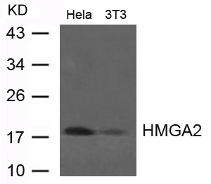
Various whole cell extracts (30 μg) were separated by 15% SDS-PAGE, and the membrane was blotted with HMGA2 antibody [HL1936] (GTX637773) diluted at 1:500. The HRP-conjugated anti-rabbit IgG antibody (GTX213110-01) was used to detect the primary antibody.
![Whole cell extract (30 μg) was separated by 15% SDS-PAGE, and the membrane was blotted with HMGA2 antibody [HL1936] (GTX637773) diluted at 1:500. The HRP-conjugated anti-rabbit IgG antibody (GTX213110-01) was used to detect the primary antibody.](https://www.genetex.com/upload/website/prouct_img/normal/GTX637773/GTX637773_T-44844_20221125_WB_R_22112723_904.webp "Whole cell extract (30 μg) was separated by 15% SDS-PAGE, and the membrane was blotted with HMGA2 antibody [HL1936] (GTX637773) diluted at 1:500. The HRP-conjugated anti-rabbit IgG antibody (GTX213110-01) was used to detect the primary antibody.")
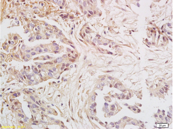
![HMGA2 antibody [HL1936] detects HMGA2 protein at nucleus by immunohistochemical analysis. Sample: Paraffin-embedded mouse stomach. HMGA2 stained by HMGA2 antibody [HL1936] (GTX637773) diluted at 1:100. Antigen Retrieval: Citrate buffer, pH 6.0, 15 min](https://www.genetex.com/upload/website/prouct_img/normal/GTX637773/GTX637773_T-44844_20221028_IHC-P_M_22122722_144.webp "HMGA2 antibody [HL1936] detects HMGA2 protein at nucleus by immunohistochemical analysis. Sample: Paraffin-embedded mouse stomach. HMGA2 stained by HMGA2 antibody [HL1936] (GTX637773) diluted at 1:100. Antigen Retrieval: Citrate buffer, pH 6.0, 15 min")
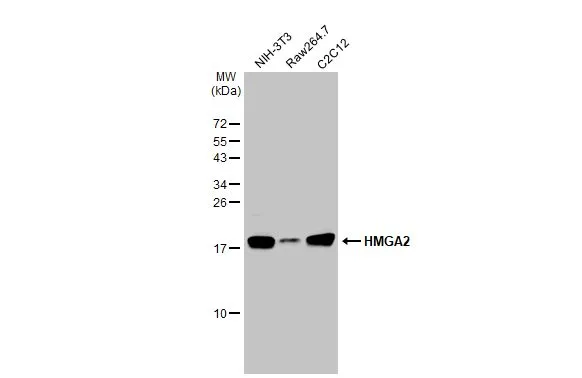
![Various whole cell extracts (30 μg) were separated by 15% SDS-PAGE, and the membrane was blotted with HMGA2 antibody [HL1936] (GTX637773) diluted at 1:500. The HRP-conjugated anti-rabbit IgG antibody (GTX213110-01) was used to detect the primary antibody. Corresponding RNA expression data for the same cell lines are based on Human Protein Atlas program.](https://www.genetex.com/upload/website/prouct_img/normal/GTX637773/GTX637773_44907_20221230_WB_TPM_watermark_23010400_176.webp "Various whole cell extracts (30 μg) were separated by 15% SDS-PAGE, and the membrane was blotted with HMGA2 antibody [HL1936] (GTX637773) diluted at 1:500. The HRP-conjugated anti-rabbit IgG antibody (GTX213110-01) was used to detect the primary antibody. Corresponding RNA expression data for the same cell lines are based on Human Protein Atlas program.")
Various whole cell extracts (30 μg) were separated by 15% SDS-PAGE, and the membrane was blotted with HMGA2 antibody [HL1936] (GTX637773) diluted at 1:500. The HRP-conjugated anti-rabbit IgG antibody (GTX213110-01) was used to detect the primary antibody.
HMGA2 antibody [HL1936]
GTX637773
ApplicationsWestern Blot, ImmunoHistoChemistry, ImmunoHistoChemistry Paraffin
Product group Antibodies
ReactivityHuman, Mouse, Rat
TargetHMGA2
Overview
- SupplierGeneTex
- Product NameHMGA2 antibody [HL1936]
- Delivery Days Customer9
- Application Supplier NoteWB: 1:500-1:3000. *Optimal dilutions/concentrations should be determined by the researcher.Not tested in other applications.
- ApplicationsWestern Blot, ImmunoHistoChemistry, ImmunoHistoChemistry Paraffin
- CertificationResearch Use Only
- ClonalityMonoclonal
- Clone IDHL1936
- Concentration1 mg/ml
- ConjugateUnconjugated
- Gene ID8091
- Target nameHMGA2
- Target descriptionhigh mobility group AT-hook 2
- Target synonymsBABL, HMGI-C, HMGIC, LIPO, SRS5, STQTL9, high mobility group protein HMGI-C, HMGA2/KRT121P fusion
- HostRabbit
- IsotypeIgG
- Protein IDP52926
- Protein NameHigh mobility group protein HMGI-C
- Scientific DescriptionThis gene encodes a protein that belongs to the non-histone chromosomal high mobility group (HMG) protein family. HMG proteins function as architectural factors and are essential components of the enhancesome. This protein contains structural DNA-binding domains and may act as a transcriptional regulating factor. Identification of the deletion, amplification, and rearrangement of this gene that are associated with myxoid liposarcoma suggests a role in adipogenesis and mesenchymal differentiation. A gene knock out study of the mouse counterpart demonstrated that this gene is involved in diet-induced obesity. Alternate transcriptional splice variants, encoding different isoforms, have been characterized. [provided by RefSeq, Jul 2008]
- ReactivityHuman, Mouse, Rat
- Storage Instruction-20°C or -80°C,2°C to 8°C
- UNSPSC41116161
Datasheet
Related products
Product group Antibodies
Anti-Hmga2 (C-term) Antibody102-24095
ApplicationsImmunoFluorescence, Western Blot
TargetHMGA2
- SizePrice
Product group Antibodies
Anti-HMGA2 AntibodyA42614
ApplicationsWestern Blot
ReactivityHuman, Mouse, Rat
- SizePrice

Product group Antibodies
Anti-HMGA2 Antibody (C-term)A00436-1
ApplicationsFlow Cytometry, Western Blot
ReactivityHuman, Mouse
TargetHMGA2
- SizePrice
Product group Antibodies
References
HMGA2 Polyclonal AntibodyBS-0556R
ApplicationsImmunoFluorescence, Western Blot, ELISA, ImmunoCytoChemistry, ImmunoHistoChemistry, ImmunoHistoChemistry Frozen, ImmunoHistoChemistry Paraffin
ReactivityChicken, Human, Mouse, Rat
TargetHMGA2
- SizePrice
Product group Antibodies

Goat anti-HMGI-C / HMGA2EB09623
ApplicationsWestern Blot, ELISA
ReactivityCanine, Human
TargetHMGA2
- SizePrice

Product group Antibodies
HMGA2 AntibodyCSB-PA701516
ApplicationsWestern Blot, ELISA
ReactivityHuman, Mouse
TargetHMGA2
- SizePrice
![HMGA2 antibody - ChIP grade detects HMGA2 protein at nucleus by immunofluorescent analysis. Sample: HeLa cells were fixed in 4% paraformaldehyde at RT for 15 min. Green: HMGA2 stained by HMGA2 antibody - ChIP grade (GTX100519) diluted at 1:500. Red: alpha Tubulin, a cytoskeleton marker, stained by alpha Tubulin antibody [GT114] (GTX628802) diluted at 1:1000.](https://www.genetex.com/upload/website/prouct_img/normal/GTX100519/GTX100519_44825_20221104_ICC_IF_22112219_632.webp)
Product group Antibodies
HMGA2 antibody - ChIP gradeGTX100519
ApplicationsImmunoFluorescence, ImmunoPrecipitation, Western Blot, ChIP Chromatin ImmunoPrecipitation, ImmunoCytoChemistry, ImmunoHistoChemistry, ImmunoHistoChemistry Paraffin
ReactivityHuman, Mouse, Rat
TargetHMGA2
- SizePrice
Product group Antibodies
HMGA2 antibody, InternalGTX88326
ApplicationsWestern Blot
ReactivityHuman
TargetHMGA2
- SizePrice
![IHC-P analysis of human follicular thyroid carcinoma (FTC) tissue using GTX639938 HMGA2 antibody [HMV314] HistoMAX?. Follicular carcinoma with strong HMGA2 staining of all tumor cells.](https://www.genetex.com/upload/website/prouct_img/normal/GTX639938/GTX639938_20240403_IHC-P_2_24040301_386.webp)
Product group Antibodies
HMGA2 antibody [HMV314] HistoMAX(tm)GTX639938
ApplicationsImmunoHistoChemistry, ImmunoHistoChemistry Paraffin
ReactivityHuman
TargetHMGA2
- SizePrice
![HMGA2 antibody [GT763] detects HMGA2 protein at nucleus by immunohistochemical analysis. Sample: Paraffin-embedded mouse brain. HMGA2 stained by HMGA2 antibody [GT763] (GTX629478) diluted at 1:200. Antigen Retrieval: Citrate buffer, pH 6.0, 15 min](https://www.genetex.com/upload/website/prouct_img/normal/GTX629478/GTX629478_43010_20181130_IHC-P_M_w_23061202_449.webp)
Product group Antibodies
HMGA2 antibody [GT763]GTX629478
ApplicationsImmunoFluorescence, ImmunoPrecipitation, Western Blot, ImmunoCytoChemistry, ImmunoHistoChemistry, ImmunoHistoChemistry Paraffin
ReactivityHuman, Mouse, Rat
TargetHMGA2
- SizePrice