

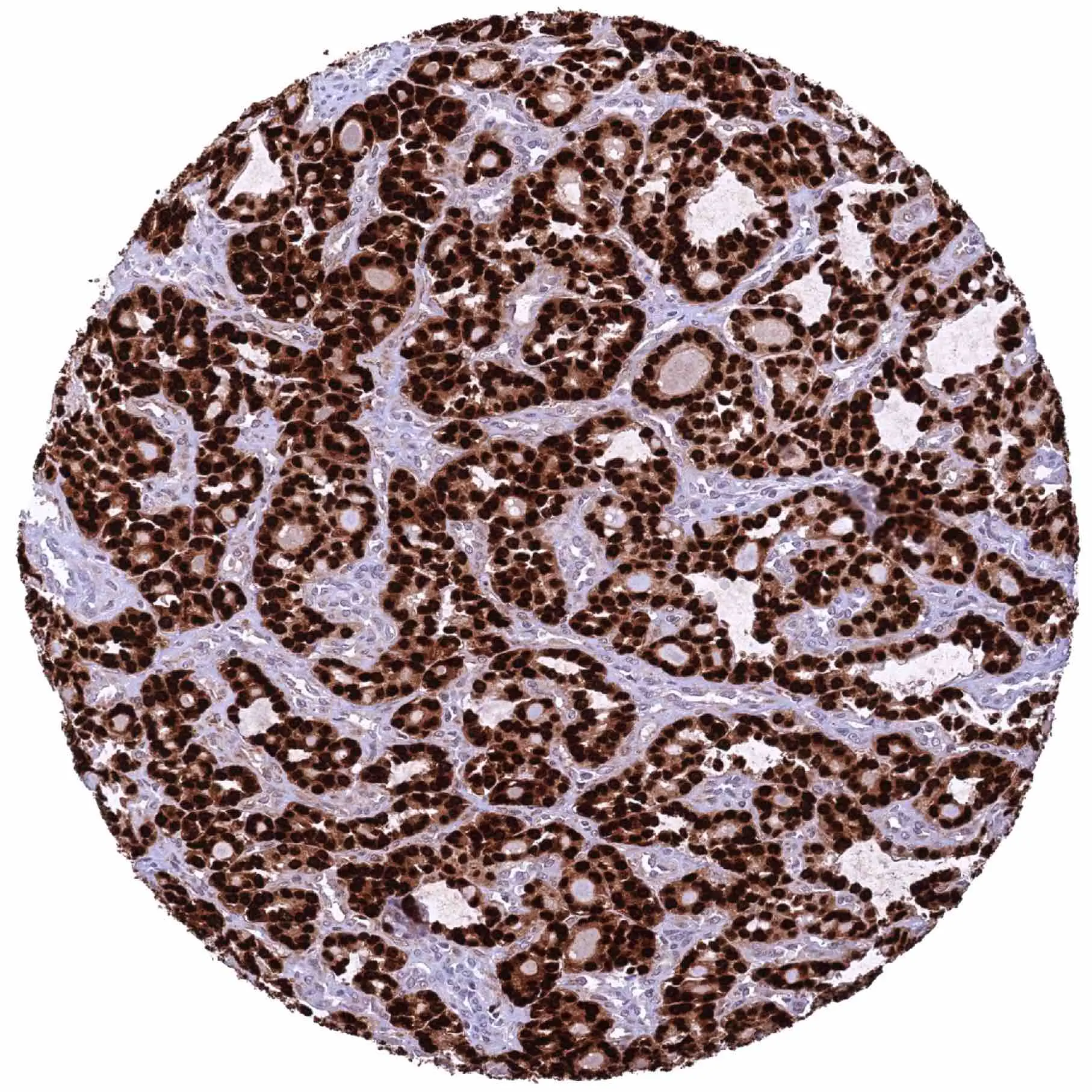
IHC-P analysis of human follicular thyroid carcinoma (FTC) tissue using GTX639938 HMGA2 antibody [HMV314] HistoMAX?. Follicular carcinoma with strong HMGA2 staining of all tumor cells.
![IHC-P analysis of human endocervix tissue using GTX639938 HMGA2 antibody [HMV314] HistoMAX?. Strong nuclear HMGA2 staining of epithelial cells.](https://www.genetex.com/upload/website/prouct_img/normal/GTX639938/GTX639938_20240403_IHC-P_24040301_329.webp "IHC-P analysis of human endocervix tissue using GTX639938 HMGA2 antibody [HMV314] HistoMAX?. Strong nuclear HMGA2 staining of epithelial cells.")
![IHC-P analysis of human bronchial mucosa tissue using GTX639938 HMGA2 antibody [HMV314] HistoMAX?. Strong nuclear HMGA2 staining of most respiratory epithelial cells.](https://www.genetex.com/upload/website/prouct_img/normal/GTX639938/GTX639938_20240403_IHC-P_1_24040301_761.webp "IHC-P analysis of human bronchial mucosa tissue using GTX639938 HMGA2 antibody [HMV314] HistoMAX?. Strong nuclear HMGA2 staining of most respiratory epithelial cells.")
![IHC-P analysis of human thyroid gland tissue using GTX639938 HMGA2 antibody [HMV314] HistoMAX?. A faint nuclear HMGA2 staining of most follicular cells.](https://www.genetex.com/upload/website/prouct_img/normal/GTX639938/GTX639938_20250214_IHC-P_1_25021323_826.webp "IHC-P analysis of human thyroid gland tissue using GTX639938 HMGA2 antibody [HMV314] HistoMAX?. A faint nuclear HMGA2 staining of most follicular cells.")
![IHC-P analysis of first-trimester human placenta tissue using GTX639938 HMGA2 antibody [HMV314] HistoMAX?. A strong nuclear HMGA2 staining of stroma cells while trophoblast cells remain HMGA2 negative.](https://www.genetex.com/upload/website/prouct_img/normal/GTX639938/GTX639938_20250214_IHC-P_2_25021323_154.webp "IHC-P analysis of first-trimester human placenta tissue using GTX639938 HMGA2 antibody [HMV314] HistoMAX?. A strong nuclear HMGA2 staining of stroma cells while trophoblast cells remain HMGA2 negative.")
![IHC-P analysis of human follicular thyroid carcinoma (FTC) tissue using GTX639938 HMGA2 antibody [HMV314] HistoMAX?. A strong nuclear HMGA2 staining of all tumor cells.](https://www.genetex.com/upload/website/prouct_img/normal/GTX639938/GTX639938_20250214_IHC-P_25021323_946.webp "IHC-P analysis of human follicular thyroid carcinoma (FTC) tissue using GTX639938 HMGA2 antibody [HMV314] HistoMAX?. A strong nuclear HMGA2 staining of all tumor cells.")
IHC-P analysis of human follicular thyroid carcinoma (FTC) tissue using GTX639938 HMGA2 antibody [HMV314] HistoMAX?. Follicular carcinoma with strong HMGA2 staining of all tumor cells.
HMGA2 antibody [HMV314] HistoMAX(tm)
GTX639938
ApplicationsImmunoHistoChemistry, ImmunoHistoChemistry Paraffin
Product group Antibodies
ReactivityHuman
TargetHMGA2
Overview
- SupplierGeneTex
- Product NameHMGA2 antibody [HMV314] HistoMAX(tm)
- Delivery Days Customer7
- Application Supplier NoteIHC-P: 1:100-1:200. *Optimal dilutions/concentrations should be determined by the researcher.Not tested in other applications.
- ApplicationsImmunoHistoChemistry, ImmunoHistoChemistry Paraffin
- CertificationResearch Use Only
- ClonalityMonoclonal
- Clone IDHMV314
- Concentration0.44 mg/ml
- ConjugateUnconjugated
- Gene ID8091
- Target nameHMGA2
- Target descriptionhigh mobility group AT-hook 2
- Target synonymsBABL, HMGI-C, HMGIC, LIPO, SRS5, STQTL9, high mobility group protein HMGI-C, HMGA2/KRT121P fusion
- HostRabbit
- IsotypeIgG
- Protein IDP52926
- Protein NameHigh mobility group protein HMGI-C
- Scientific DescriptionThis gene encodes a protein that belongs to the non-histone chromosomal high mobility group (HMG) protein family. HMG proteins function as architectural factors and are essential components of the enhancesome. This protein contains structural DNA-binding domains and may act as a transcriptional regulating factor. Identification of the deletion, amplification, and rearrangement of this gene that are associated with myxoid liposarcoma suggests a role in adipogenesis and mesenchymal differentiation. A gene knock out study of the mouse counterpart demonstrated that this gene is involved in diet-induced obesity. Alternate transcriptional splice variants, encoding different isoforms, have been characterized. [provided by RefSeq, Jul 2008]
- ReactivityHuman
- Storage Instruction-20°C or -80°C,2°C to 8°C
- UNSPSC41116161
Datasheet
Related products
Product group Antibodies
Anti-Hmga2 (C-term) Antibody102-24095
ApplicationsImmunoFluorescence, Western Blot
TargetHMGA2
- SizePrice
Product group Antibodies
Anti-HMGA2 AntibodyA42614
ApplicationsWestern Blot
ReactivityHuman, Mouse, Rat
- SizePrice

Product group Antibodies
Anti-HMGA2 Antibody (C-term)A00436-1
ApplicationsFlow Cytometry, Western Blot
ReactivityHuman, Mouse
TargetHMGA2
- SizePrice
Product group Antibodies
References
HMGA2 Polyclonal AntibodyBS-0556R
ApplicationsImmunoFluorescence, Western Blot, ELISA, ImmunoCytoChemistry, ImmunoHistoChemistry, ImmunoHistoChemistry Frozen, ImmunoHistoChemistry Paraffin
ReactivityChicken, Human, Mouse, Rat
TargetHMGA2
- SizePrice
Product group Antibodies

Goat anti-HMGI-C / HMGA2EB09623
ApplicationsWestern Blot, ELISA
ReactivityCanine, Human
TargetHMGA2
- SizePrice

Product group Antibodies
HMGA2 AntibodyCSB-PA701516
ApplicationsWestern Blot, ELISA
ReactivityHuman, Mouse
TargetHMGA2
- SizePrice
![HMGA2 antibody - ChIP grade detects HMGA2 protein at nucleus by immunofluorescent analysis. Sample: HeLa cells were fixed in 4% paraformaldehyde at RT for 15 min. Green: HMGA2 stained by HMGA2 antibody - ChIP grade (GTX100519) diluted at 1:500. Red: alpha Tubulin, a cytoskeleton marker, stained by alpha Tubulin antibody [GT114] (GTX628802) diluted at 1:1000.](https://www.genetex.com/upload/website/prouct_img/normal/GTX100519/GTX100519_44825_20221104_ICC_IF_22112219_632.webp)
Product group Antibodies
HMGA2 antibody - ChIP gradeGTX100519
ApplicationsImmunoFluorescence, ImmunoPrecipitation, Western Blot, ChIP Chromatin ImmunoPrecipitation, ImmunoCytoChemistry, ImmunoHistoChemistry, ImmunoHistoChemistry Paraffin
ReactivityHuman, Mouse, Rat
TargetHMGA2
- SizePrice
Product group Antibodies
HMGA2 antibody, InternalGTX88326
ApplicationsWestern Blot
ReactivityHuman
TargetHMGA2
- SizePrice
![Various whole cell extracts (30 μg) were separated by 15% SDS-PAGE, and the membrane was blotted with HMGA2 antibody [HL1936] (GTX637773) diluted at 1:500. The HRP-conjugated anti-rabbit IgG antibody (GTX213110-01) was used to detect the primary antibody.](https://www.genetex.com/upload/website/prouct_img/normal/GTX637773/GTX637773_T-44844_20221125_WB_M_22112723_710.webp)
Product group Antibodies
HMGA2 antibody [HL1936]GTX637773
ApplicationsWestern Blot, ImmunoHistoChemistry, ImmunoHistoChemistry Paraffin
ReactivityHuman, Mouse, Rat
TargetHMGA2
- SizePrice
![HMGA2 antibody [GT763] detects HMGA2 protein at nucleus by immunohistochemical analysis. Sample: Paraffin-embedded mouse brain. HMGA2 stained by HMGA2 antibody [GT763] (GTX629478) diluted at 1:200. Antigen Retrieval: Citrate buffer, pH 6.0, 15 min](https://www.genetex.com/upload/website/prouct_img/normal/GTX629478/GTX629478_43010_20181130_IHC-P_M_w_23061202_449.webp)
Product group Antibodies
HMGA2 antibody [GT763]GTX629478
ApplicationsImmunoFluorescence, ImmunoPrecipitation, Western Blot, ImmunoCytoChemistry, ImmunoHistoChemistry, ImmunoHistoChemistry Paraffin
ReactivityHuman, Mouse, Rat
TargetHMGA2
- SizePrice