

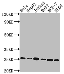
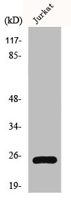
Western Blot Positive WB detected in: Hela whole cell lysate, HepG2 whole cell lysate, Jurkat whole cell lysate, 293T whole cell lysate, MCF-7 whole cell lysate, HL60 whole cell lysate All lanes: HMGB1 antibody at 1:2000 Secondary Goat polyclonal to rabbit IgG at 1/50000 dilution Predicted band size: 25 kDa Observed band size: 25 kDa
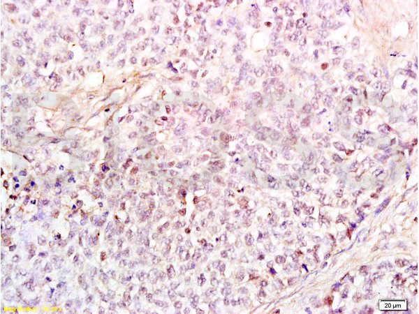
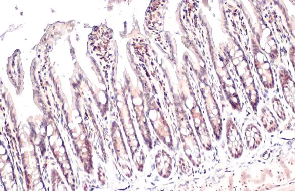
. Section was blocked with 10% normal goat serum 30min at RT. Then primary antibody (1% BSA) was incubated at 4°C overnight. The primary is detected by a Goat anti-rabbit IgG polymer labeled by HRP and visualized using 0.05% DAB.")
Western Blot Positive WB detected in: Hela whole cell lysate, HepG2 whole cell lysate, Jurkat whole cell lysate, 293T whole cell lysate, MCF-7 whole cell lysate, HL60 whole cell lysate All lanes: HMGB1 antibody at 1:2000 Secondary Goat polyclonal to rabbit IgG at 1/50000 dilution Predicted band size: 25 kDa Observed band size: 25 kDa
HMGB1 Recombinant Monoclonal Antibody
CSB-RA439052A0HU
ApplicationsWestern Blot, ELISA, ImmunoHistoChemistry
Product group Antibodies
ReactivityHuman
TargetHMGB1
Overview
- SupplierCusabio
- Product NameHMGB1 Recombinant Monoclonal Antibody
- Delivery Days Customer20
- ApplicationsWestern Blot, ELISA, ImmunoHistoChemistry
- CertificationResearch Use Only
- ClonalityMonoclonal
- Clone ID1A1
- ConjugateUnconjugated
- Gene ID3146
- Target nameHMGB1
- Target descriptionhigh mobility group box 1
- Target synonymsHMG-1, HMG1, HMG3, SBP-1, high mobility group protein B1, Amphoterin, Sulfoglucuronyl carbohydrate binding protein, high-mobility group (nonhistone chromosomal) protein 1
- IsotypeIgG
- Protein IDP09429
- Protein NameHigh mobility group protein B1
- Scientific DescriptionMultifunctional redox sensitive protein with various roles in different cellular compartments. In the nucleus is one of the major chromatin-associated non-histone proteins and acts as a DNA chaperone involved in replication, transcription, chromatin remodeling, V(D)J recombination, DNA repair and genome stability. Proposed to be an universal biosensor for nucleic acids. Promotes host inflammatory response to sterile and infectious signals and is involved in the coordination and integration of innate and adaptive immune responses. In the cytoplasm functions as sensor and/or chaperone for immunogenic nucleic acids implicating the activation of TLR9-mediated immune responses, and mediates autophagy. Acts as danger associated molecular pattern (DAMP) molecule that amplifies immune responses during tissue injury (PubMed:27362237). Released to the extracellular environment can bind DNA, nucleosomes, IL-1 beta, CXCL12, AGER isoform 2/sRAGE, lipopolysaccharide (LPS) and lipoteichoic acid (LTA), and activates cells through engagement of multiple surface receptors. In the extracellular compartment fully reduced HMGB1 (released by necrosis) acts as a chemokine, disulfide HMGB1 (actively secreted) as a cytokine, and sulfonyl HMGB1 (released from apoptotic cells) promotes immunological tolerance (PubMed:23519706, PubMed:23446148, PubMed:23994764, PubMed:25048472). Has proangiogdenic activity (By similarity). May be involved in platelet activation (By similarity). Binds to phosphatidylserine and phosphatidylethanolamide (By similarity). Bound to RAGE mediates signaling for neuronal outgrowth (By similarity). May play a role in accumulation of expanded polyglutamine (polyQ) proteins such as huntingtin (HTT) or TBP (PubMed:23303669, PubMed:25549101).
- ReactivityHuman
- Storage Instruction-20°C or -80°C
- UNSPSC41116161
Related products
Product group Antibodies
Anti-HMGB1 AntibodyA97486
ApplicationsWestern Blot, ELISA
ReactivityHuman, Mouse, Rat
- SizePrice
Product group Antibodies
anti-HMGB1, mAb (rec.) (Giby-1-4)AG-27B-0002
ApplicationsWestern Blot, ELISA
ReactivityHuman, Mouse, Rat
TargetHMGB1
- SizePrice
Product group Antibodies
Anti-HMGB1 Antibody144-02553
ApplicationsImmunoFluorescence, Western Blot, ImmunoHistoChemistry
ReactivityHuman, Mouse, Rat
TargetHMGB1
- SizePrice

Product group Antibodies
Anti-HMGB1 Antibody Picoband(r)A00066-1-CARRIER-FREE
ApplicationsFlow Cytometry, ImmunoFluorescence, Western Blot, ImmunoCytoChemistry, ImmunoHistoChemistry
ReactivityHuman, Mouse, Rat
TargetHMGB1
- SizePrice
Product group Antibodies
References
HMGB1 Polyclonal AntibodyBS-0664R
ApplicationsFlow Cytometry, ImmunoFluorescence, Western Blot, ELISA, ImmunoCytoChemistry, ImmunoHistoChemistry, ImmunoHistoChemistry Frozen, ImmunoHistoChemistry Paraffin
ReactivityBovine, Human, Mouse, Rat
TargetHMGB1
- SizePrice
Product group Antibodies
HMGB1 AntibodyCSB-PA002937
ApplicationsImmunoFluorescence, Western Blot, ELISA, ImmunoHistoChemistry
ReactivityHuman, Mouse, Rat
TargetHMGB1
- SizePrice
Product group Antibodies
Hmgb1 Polyclonal AntibodyCAC07036
ApplicationsWestern Blot, ELISA, ImmunoHistoChemistry
ReactivityMouse
TargetHMGB1
- SizePrice
Product group Antibodies
HMGB1 antibodyGTX101277
ApplicationsImmunoFluorescence, Western Blot, ImmunoCytoChemistry, ImmunoHistoChemistry, ImmunoHistoChemistry Paraffin
ReactivityHuman, Mouse, Porcine, Rat, Zebra Fish
TargetHMGB1
- SizePrice

Product group Antibodies
Anti-HMGB1 AntibodyHPA073663
ApplicationsWestern Blot, ImmunoCytoChemistry
ReactivityHuman
TargetHMGB1
- SizePrice