

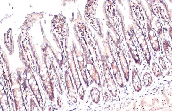
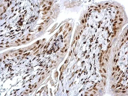
HMGB1 antibody detects HMGB1 protein at nucleus by immunohistochemical analysis. Sample: Paraffin-embedded mouse intestine. HMGB1 stained by HMGB1 antibody (GTX101277) diluted at 1:500. Antigen Retrieval: Citrate buffer, pH 6.0, 15 min
 diluted at 1:500. Antigen Retrieval: Citrate buffer, pH 6.0, 15 min")
 diluted at 1:500. Antigen Retrieval: Citrate buffer, pH 6.0, 15 min")
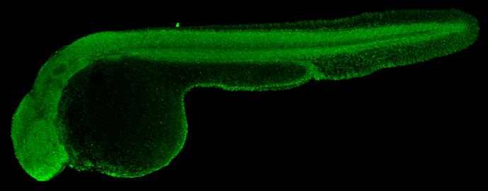
 diluted at 1:100. Antigen Retrieval: Tris-HCl buffer, pH 9.0, 20 min at 70oC")
 at 1:300 dilution.")
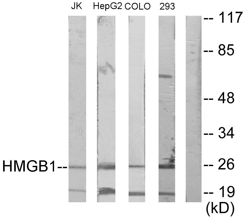
 were separated by 12% SDS-PAGE, and the membrane was blotted with HMGB1 antibody (GTX101277) diluted at 1:1000. The HRP-conjugated anti-rabbit IgG antibody (GTX213110-01) was used to detect the primary antibody. (GI: gastrointestinal)")
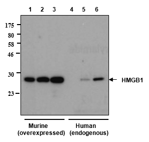
 and transfected (+) 293T whole cell extracts (30 μg) were separated by 12% SDS-PAGE, and the membrane was blotted with HMGB1 antibody (GTX101277) diluted at 1:5000. The HRP-conjugated anti-rabbit IgG antibody (GTX213110-01) was used to detect the primary antibody.")
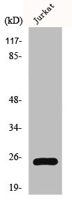
 dilution: 1:3000 The HRP-conjugated anti-rabbit IgG antibody (GTX213110-01) was used to detect the primary antibody.")
 dilution: 1:1000.
Antigen Retrieval: Trilogy? (EDTA based, pH 8.0) buffer, 15min")
 dilution: 1:1000.
Antigen Retrieval: Trilogy? (EDTA based, pH 8.0) buffer, 15min")
HMGB1 antibody detects HMGB1 protein at nucleus by immunohistochemical analysis. Sample: Paraffin-embedded mouse intestine. HMGB1 stained by HMGB1 antibody (GTX101277) diluted at 1:500. Antigen Retrieval: Citrate buffer, pH 6.0, 15 min
HMGB1 antibody
GTX101277
ApplicationsImmunoFluorescence, Western Blot, ImmunoCytoChemistry, ImmunoHistoChemistry, ImmunoHistoChemistry Paraffin
Product group Antibodies
ReactivityHuman, Mouse, Porcine, Rat, Zebra Fish
TargetHMGB1
Overview
- SupplierGeneTex
- Product NameHMGB1 antibody
- Delivery Days Customer9
- Application Supplier NoteWB: 1:500-1:3000. ICC/IF: 1:100-1:1000. IHC-P: 1:100-1:1000. *Optimal dilutions/concentrations should be determined by the researcher.Not tested in other applications.
- ApplicationsImmunoFluorescence, Western Blot, ImmunoCytoChemistry, ImmunoHistoChemistry, ImmunoHistoChemistry Paraffin
- CertificationResearch Use Only
- ClonalityPolyclonal
- Concentration0.43 mg/ml
- ConjugateUnconjugated
- Gene ID3146
- Target nameHMGB1
- Target descriptionhigh mobility group box 1
- Target synonymsHMG-1, HMG1, HMG3, SBP-1, high mobility group protein B1, Amphoterin, Sulfoglucuronyl carbohydrate binding protein, high-mobility group (nonhistone chromosomal) protein 1
- HostRabbit
- IsotypeIgG
- Protein IDP09429
- Protein NameHigh mobility group protein B1
- Scientific DescriptionDNA binding proteins that associates with chromatin and has the ability to bend DNA. Binds preferentially single-stranded DNA. Involved in V(D)J recombination by acting as a cofactor of the RAG complex. Acts by stimulating cleavage and RAG protein binding at the 23 bp spacer of conserved recombination signal sequences (RSS). Heparin-binding protein that has a role in the extension of neurite-type cytoplasmic processes in developing cells.
- ReactivityHuman, Mouse, Porcine, Rat, Zebra Fish
- Storage Instruction-20°C or -80°C,2°C to 8°C
- UNSPSC41116161
Datasheet
Related products
Product group Antibodies
Anti-HMGB1 Antibody144-02553
ApplicationsImmunoFluorescence, Western Blot, ImmunoHistoChemistry
ReactivityHuman, Mouse, Rat
TargetHMGB1
- SizePrice
Product group Antibodies
Anti-HMGB1 AntibodyA97486
ApplicationsWestern Blot, ELISA
ReactivityHuman, Mouse, Rat
- SizePrice

Product group Antibodies
Anti-HMGB1 Antibody Picoband(r)A00066-1-CARRIER-FREE
ApplicationsFlow Cytometry, ImmunoFluorescence, Western Blot, ImmunoCytoChemistry, ImmunoHistoChemistry
ReactivityHuman, Mouse, Rat
TargetHMGB1
- SizePrice
Product group Antibodies
anti-HMGB1, mAb (rec.) (Giby-1-4)AG-27B-0002
ApplicationsWestern Blot, ELISA
ReactivityHuman, Mouse, Rat
TargetHMGB1
- SizePrice
Product group Antibodies
Hmgb1 Polyclonal AntibodyCAC07036
ApplicationsWestern Blot, ELISA, ImmunoHistoChemistry
ReactivityMouse
TargetHMGB1
- SizePrice
Product group Antibodies
HMGB1 AntibodyCSB-PA002937
ApplicationsImmunoFluorescence, Western Blot, ELISA, ImmunoHistoChemistry
ReactivityHuman, Mouse, Rat
TargetHMGB1
- SizePrice
Product group Antibodies
HMGB1 antibodyGTX112959
ApplicationsImmunoFluorescence, Western Blot, ImmunoCytoChemistry, ImmunoHistoChemistry, ImmunoHistoChemistry Paraffin
ReactivityHuman, Mouse, Rat, Zebra Fish
TargetHMGB1
- SizePrice
Product group Antibodies
References
HMGB1 antibody [HAP46.5]GTX12029
ApplicationsWestern Blot, ELISA
ReactivityCanine, Chicken, Human, Mouse, Rat
TargetHMGB1
- SizePrice
Product group Antibodies
HMGB1 antibodyGTX127344
ApplicationsImmunoFluorescence, ImmunoPrecipitation, Western Blot, ImmunoCytoChemistry, ImmunoHistoChemistry, ImmunoHistoChemistry Frozen, ImmunoHistoChemistry Paraffin, Other Application
ReactivityHuman, Mouse, Rat
TargetHMGB1
- SizePrice