

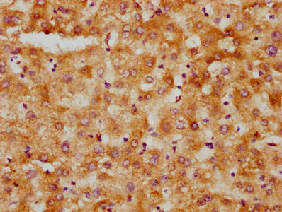
IHC image of CSB-PA010566LA01HU diluted at 1:500 and staining in paraffin-embedded human liver tissue performed on a Leica BondTM system. After dewaxing and hydration, antigen retrieval was mediated by high pressure in a citrate buffer (pH 6.0). Section was blocked with 10% normal goat serum 30min at RT. Then primary antibody (1% BSA) was incubated at 4°C overnight. The primary is detected by a biotinylated secondary antibody and visualized using an HRP conjugated SP system.
. Section was blocked with 10% normal goat serum 30min at RT. Then primary antibody (1% BSA) was incubated at 4°C overnight. The primary is detected by a biotinylated secondary antibody and visualized using an HRP conjugated SP system.")
.")
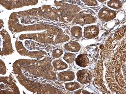
IHC image of CSB-PA010566LA01HU diluted at 1:500 and staining in paraffin-embedded human liver tissue performed on a Leica BondTM system. After dewaxing and hydration, antigen retrieval was mediated by high pressure in a citrate buffer (pH 6.0). Section was blocked with 10% normal goat serum 30min at RT. Then primary antibody (1% BSA) was incubated at 4°C overnight. The primary is detected by a biotinylated secondary antibody and visualized using an HRP conjugated SP system.
HMGCS1 Antibody
CSB-PA010566LA01HU
ApplicationsImmunoFluorescence, ELISA, ImmunoHistoChemistry
Product group Antibodies
ReactivityHuman
TargetHMGCS1
Overview
- SupplierCusabio
- Product NameHMGCS1 Antibody
- Delivery Days Customer20
- ApplicationsImmunoFluorescence, ELISA, ImmunoHistoChemistry
- CertificationResearch Use Only
- ClonalityPolyclonal
- ConjugateUnconjugated
- Gene ID3157
- Target nameHMGCS1
- Target description3-hydroxy-3-methylglutaryl-CoA synthase 1
- Target synonymsHMGCS, hydroxymethylglutaryl-CoA synthase, cytoplasmic, 3-hydroxy-3-methylglutaryl coenzyme A (HMG-CoA) synthase, 3-hydroxy-3-methylglutaryl-CoA synthase 1 (soluble), 3-hydroxy-3-methylglutaryl-Coenzyme A synthase 1 (soluble)
- HostRabbit
- IsotypeIgG
- Protein IDQ01581
- Protein NameHydroxymethylglutaryl-CoA synthase, cytoplasmic
- Scientific DescriptionThis enzyme condenses acetyl-CoA with acetoacetyl-CoA to form HMG-CoA, which is the substrate for HMG-CoA reductase.
- ReactivityHuman
- Storage Instruction-20°C or -80°C
- UNSPSC41116161
Related products

Product group Antibodies
Anti-HMGCS1 AntibodyA9326
ApplicationsImmunoFluorescence, Western Blot, ImmunoCytoChemistry
ReactivityHuman, Mouse, Rat
- SizePrice

Product group Antibodies
Anti-HMGCS1 Antibody Picoband(r)A05313-2-CARRIER-FREE
ApplicationsFlow Cytometry, ImmunoFluorescence, Western Blot, ELISA, ImmunoCytoChemistry, ImmunoHistoChemistry
ReactivityHuman
TargetHMGCS1
- SizePrice

Product group Antibodies
Anti-HMGCS1 AntibodyHPA036913
ApplicationsWestern Blot, ImmunoHistoChemistry
ReactivityHuman
TargetHMGCS1
- SizePrice
Product group Antibodies

Goat anti-HMGCS1EB12867
ApplicationsWestern Blot, ELISA
ReactivityHuman
TargetHMGCS1
- SizePrice
Product group Antibodies

HMGCS1 / HMG-CoA Synthase 1 AntibodyLS-C403210
ApplicationsWestern Blot, ELISA, ImmunoHistoChemistry
ReactivityHuman
TargetHMGCS1
- SizePrice
Product group Antibodies
HMGCS1 Polyclonal AntibodyCAC13079
ApplicationsImmunoFluorescence, ELISA, ImmunoHistoChemistry
TargetHMGCS1
- SizePrice
Product group Antibodies
HMGCS1 antibodyGTX112346
ApplicationsImmunoFluorescence, Western Blot, ImmunoCytoChemistry, ImmunoHistoChemistry, ImmunoHistoChemistry Frozen, ImmunoHistoChemistry Paraffin
ReactivityHuman, Mouse
TargetHMGCS1
- SizePrice
Product group Antibodies
Anti-HMGCS1 Antibody144-03916
ApplicationsWestern Blot
ReactivityHuman, Mouse, Rat
TargetHMGCS1
- SizePrice