

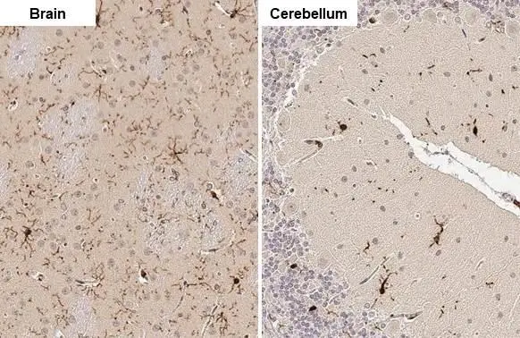
Iba1 antibody detects Iba1 protein by immunohistochemical analysis. Sample: Paraffin-embedded rat tissues. Iba1 stained by Iba1 antibody (GTX100042) diluted at 1:100. Antigen Retrieval: Citrate buffer, pH 6.0, 15 min
 diluted at 1:500. Blue: Fluoroshield with DAPI (GTX30920).")
 were separated by 15% SDS-PAGE, and the membrane was blotted with Iba1 antibody (GTX100042) diluted at 1:1000. The HRP-conjugated anti-rabbit IgG antibody (GTX213110-01) was used to detect the primary antibody.")
 diluted at 1:500. Antigen Retrieval: Citrate buffer, pH 6.0, 15 min")
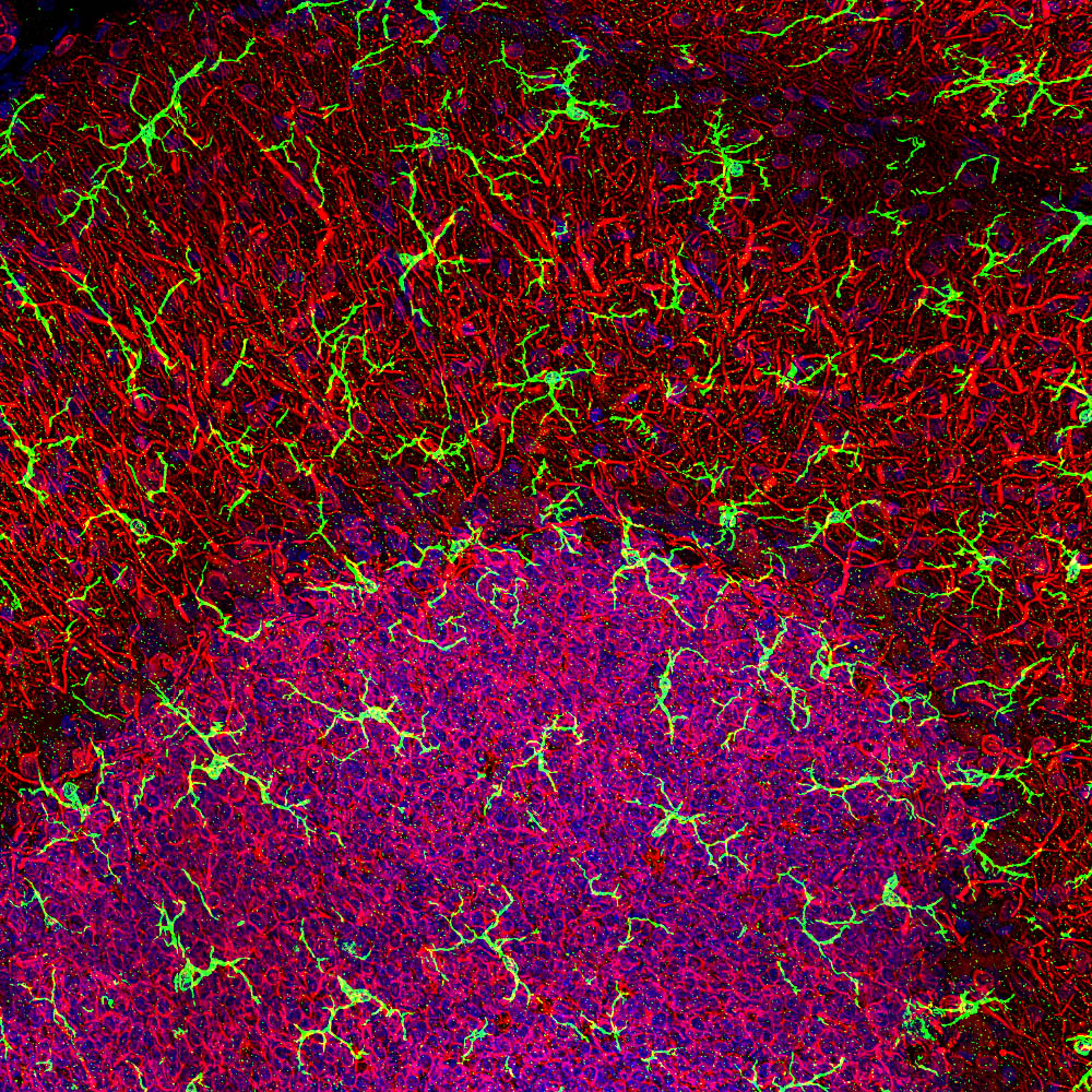
![Iba1 antibody detects Iba1 protein expression at microglias by immunohistochemical analysis. Sample: Frozen sectioned E13.5 Rat brain. Green: Iba1 protein stained by Iba1 antibody (GTX100042) diluted at 1:250. Red: beta Tubulin 3/ TUJ1, a mature neuron marker, stained by beta Tubulin 3/ TUJ1 antibody [GT11710] (GTX631836) diluted at 1:500. Blue: Fluoroshield with DAPI (GTX30920).](https://www.genetex.com/upload/website/prouct_img/normal/GTX100042/GTX100042_41556_20161005_IHC-Fr_R_w_23053123_599.webp "Iba1 antibody detects Iba1 protein expression at microglias by immunohistochemical analysis. Sample: Frozen sectioned E13.5 Rat brain. Green: Iba1 protein stained by Iba1 antibody (GTX100042) diluted at 1:250. Red: beta Tubulin 3/ TUJ1, a mature neuron marker, stained by beta Tubulin 3/ TUJ1 antibody [GT11710] (GTX631836) diluted at 1:500. Blue: Fluoroshield with DAPI (GTX30920).")
 diluted at 1:500. Blue: Fluoroshield with DAPI (GTX30920). Antigen Retrieval: ice-cold MeOH for 5 min")
 diluted at 1:500. Antigen Retrieval: Citrate buffer, pH 6.0, 15 min")
 was separated by 15% SDS-PAGE, and the membrane was blotted with Iba1 antibody (GTX100042) diluted at 1:1000.")
 dilution: 1:500.
Antigen Retrieval: Trilogy? (EDTA based, pH 8.0) buffer, 15min")
 diluted at 1:500. Antigen Retrieval: Citrate buffer, pH 6.0, 15 min")
Iba1 antibody detects Iba1 protein by immunohistochemical analysis. Sample: Paraffin-embedded rat tissues. Iba1 stained by Iba1 antibody (GTX100042) diluted at 1:100. Antigen Retrieval: Citrate buffer, pH 6.0, 15 min
Iba1 antibody
GTX100042
ApplicationsFlow Cytometry, ImmunoFluorescence, Western Blot, ImmunoCytoChemistry, ImmunoHistoChemistry, ImmunoHistoChemistry Frozen, ImmunoHistoChemistry Paraffin
Product group Antibodies
ReactivityHuman, Mouse, Rat
TargetAIF1
Overview
- SupplierGeneTex
- Product NameIba1 antibody
- Delivery Days Customer9
- Application Supplier NoteWB: 1:500-1:10000. ICC/IF: 1:100-1:1000. IHC-P: 1:100-1:1000. IHC-Fr: 1:100-1:1000. FCM: 1:50-1:200. *Optimal dilutions/concentrations should be determined by the researcher.Not tested in other applications.
- ApplicationsFlow Cytometry, ImmunoFluorescence, Western Blot, ImmunoCytoChemistry, ImmunoHistoChemistry, ImmunoHistoChemistry Frozen, ImmunoHistoChemistry Paraffin
- CertificationResearch Use Only
- ClonalityPolyclonal
- Concentration1 mg/ml
- ConjugateUnconjugated
- Gene ID199
- Target nameAIF1
- Target descriptionallograft inflammatory factor 1
- Target synonymsAIF-1, IBA1, IRT-1, IRT1, allograft inflammatory factor 1, interferon gamma responsive transcript, ionized calcium-binding adapter molecule 1, protein G1
- HostRabbit
- IsotypeIgG
- Protein IDP55008
- Protein NameAllograft inflammatory factor 1
- Scientific DescriptionThis gene is induced by cytokines and interferon. Its protein product is thought to be involved in negative regulation of growth of vascular smooth muscle cells, which contributes to the anti-inflammatory response to vessel wall trauma. Three transcript variants encoding different isoforms have been found for this gene. [provided by RefSeq]
- ReactivityHuman, Mouse, Rat
- Storage Instruction-20°C or -80°C,2°C to 8°C
- UNSPSC41116161
Datasheet
Related products
Product group Antibodies
Anti-Iba1 AntibodyA104332
ApplicationsImmunoFluorescence, Western Blot, ImmunoCytoChemistry, ImmunoHistoChemistry
ReactivityHuman, Mouse, Rat
- SizePrice
Product group Antibodies
Anti-AIF1 Antibody144-60080
ApplicationsWestern Blot
ReactivityHuman, Mouse, Rat
TargetAIF1
- SizePrice
Product group Antibodies

Anti-AIF1 [GT1-mAb1]AB03986-1.1
ApplicationsFlow Cytometry, ImmunoFluorescence, Western Blot, ELISA
ReactivityHuman
TargetAIF1
- SizePrice

Product group Antibodies
Anti-AIF1 AntibodyAMAB91671
ApplicationsImmunoHistoChemistry
ReactivityHuman
TargetAIF1
- SizePrice
Product group Antibodies

AIF1 / IBA1 AntibodyLS-C763926
ApplicationsELISA, ImmunoHistoChemistry
ReactivityHuman, Mouse, Rat
TargetAIF1
- SizePrice

Product group Antibodies
Anti-Iba1/AIF1 Antibody Picoband(r)A01394-CARRIER-FREE
ApplicationsWestern Blot, ImmunoHistoChemistry
ReactivityHuman
TargetAIF1
- SizePrice
Product group Antibodies
References
AIF1/Iba1 Polyclonal AntibodyBS-1363R
ApplicationsFlow Cytometry, ImmunoFluorescence, Western Blot, ELISA, ImmunoCytoChemistry, ImmunoHistoChemistry, ImmunoHistoChemistry Frozen, ImmunoHistoChemistry Paraffin
ReactivityHuman, Mouse, Rat
TargetAIF1
- SizePrice
Product group Antibodies
AIF1 AntibodyCSB-PA00667A0RB
ApplicationsImmunoFluorescence, ELISA, ImmunoHistoChemistry
ReactivityHuman
TargetAIF1
- SizePrice
Product group Antibodies

ApplicationsWestern Blot, ELISA
ReactivityHuman, Mouse, Porcine, Rat
TargetAIF1
- SizePrice