

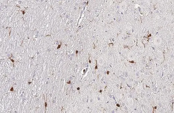
Iba1 antibody [HL1880-MS] detects Iba 1 protein by immunohistochemical analysis. Sample: Paraffin-embedded rat brain. Green: Iba 1 stained by Iba1 antibody [HL1880-MS] (GTX638147) diluted at 1:100. Antigen Retrieval: Citrate buffer, pH 6.0, 15 min
![Iba1 antibody [HL1880-MS] detects Iba 1 protein by immunohistochemical analysis. Sample: Paraffin-embedded mouse brain. Green: Iba 1 stained by Iba1 antibody [HL1880-MS] (GTX638147) diluted at 1:100. Antigen Retrieval: Citrate buffer, pH 6.0, 15 min](https://www.genetex.com/upload/website/prouct_img/normal/GTX638147/GTX638147_T-44928_20230120_IHC-P_M_23020621_536.webp "Iba1 antibody [HL1880-MS] detects Iba 1 protein by immunohistochemical analysis. Sample: Paraffin-embedded mouse brain. Green: Iba 1 stained by Iba1 antibody [HL1880-MS] (GTX638147) diluted at 1:100. Antigen Retrieval: Citrate buffer, pH 6.0, 15 min")
![Iba1 antibody [HL1880-MS] detects Iba1 protein at cell membrane by immunofluorescent analysis. Sample: THP-1 cells were fixed in 4% paraformaldehyde at RT for 15 min. Green: Iba1 stained by Iba1 antibody [HL1880-MS] (GTX638147) diluted at 1:500. Blue: Fluoroshield with DAPI (GTX30920).](https://www.genetex.com/upload/website/prouct_img/normal/GTX638147/GTX638147_T-44928_20230331_ICC_IF_23041023_532.webp "Iba1 antibody [HL1880-MS] detects Iba1 protein at cell membrane by immunofluorescent analysis. Sample: THP-1 cells were fixed in 4% paraformaldehyde at RT for 15 min. Green: Iba1 stained by Iba1 antibody [HL1880-MS] (GTX638147) diluted at 1:500. Blue: Fluoroshield with DAPI (GTX30920).")
![Iba1 antibody [HL1880-MS] detects Iba1 protein at cell membrane and cytoplasm by immunohistochemical analysis. Sample: Frozen-sectioned mouse brain. Green: Iba1 stained by Iba1 antibody [HL1880-MS] (GTX638147) diluted at 1:100. Blue: Fluoroshield with DAPI (GTX30920).](https://www.genetex.com/upload/website/prouct_img/normal/GTX638147/GTX638147_T-44928_20230414_IHC-Fr_M_23041719_233.webp "Iba1 antibody [HL1880-MS] detects Iba1 protein at cell membrane and cytoplasm by immunohistochemical analysis. Sample: Frozen-sectioned mouse brain. Green: Iba1 stained by Iba1 antibody [HL1880-MS] (GTX638147) diluted at 1:100. Blue: Fluoroshield with DAPI (GTX30920).")
![Iba1 antibody [HL1880-MS] detects Iba1 protein by immunohistochemical analysis. Sample: Frozen-sectioned mouse spinal cord. Green: Iba1 stained by Iba1 antibody [HL1880-MS] (GTX638147) diluted at 1:50. Blue: Fluoroshield with DAPI (GTX30920).](https://www.genetex.com/upload/website/prouct_img/normal/GTX638147/GTX638147_45033_20230630_IHC-Fr_M_23071822_688.webp "Iba1 antibody [HL1880-MS] detects Iba1 protein by immunohistochemical analysis. Sample: Frozen-sectioned mouse spinal cord. Green: Iba1 stained by Iba1 antibody [HL1880-MS] (GTX638147) diluted at 1:50. Blue: Fluoroshield with DAPI (GTX30920).")
![Iba1 antibody [HL1880-MS] detects Iba1 protein by immunohistochemical analysis. Sample: Paraffin-embedded rat brain. Green: Iba1 stained by Iba1 antibody [HL1880-MS] (GTX638147) diluted at 1:250. Blue: Hoechst 33342 staining. Antigen Retrieval: Citrate buffer, pH 6.0, 15 min](https://www.genetex.com/upload/website/prouct_img/normal/GTX638147/GTX638147_45201_20240626_IHC-P_R_24070822_707.webp "Iba1 antibody [HL1880-MS] detects Iba1 protein by immunohistochemical analysis. Sample: Paraffin-embedded rat brain. Green: Iba1 stained by Iba1 antibody [HL1880-MS] (GTX638147) diluted at 1:250. Blue: Hoechst 33342 staining. Antigen Retrieval: Citrate buffer, pH 6.0, 15 min")
![Various whole cell extracts (30 μg) were separated by 15% SDS-PAGE, and the membrane was blotted with Iba1 antibody [HL1880-MS] (GTX638147) diluted at 1:1000. The HRP-conjugated anti-mouse IgG antibody (GTX213111-01) was used to detect the primary antibody. Corresponding RNA expression data for the same cell lines are based on Human Protein Atlas program.](https://www.genetex.com/upload/website/prouct_img/normal/GTX638147/GTX638147_45481_20240726_WB_TPM_watermark_24103022_871.webp "Various whole cell extracts (30 μg) were separated by 15% SDS-PAGE, and the membrane was blotted with Iba1 antibody [HL1880-MS] (GTX638147) diluted at 1:1000. The HRP-conjugated anti-mouse IgG antibody (GTX213111-01) was used to detect the primary antibody. Corresponding RNA expression data for the same cell lines are based on Human Protein Atlas program.")
Iba1 antibody [HL1880-MS] detects Iba 1 protein by immunohistochemical analysis. Sample: Paraffin-embedded rat brain. Green: Iba 1 stained by Iba1 antibody [HL1880-MS] (GTX638147) diluted at 1:100. Antigen Retrieval: Citrate buffer, pH 6.0, 15 min
Iba1 antibody [HL1880-MS]
GTX638147
ApplicationsImmunoFluorescence, Western Blot, ImmunoCytoChemistry, ImmunoHistoChemistry, ImmunoHistoChemistry Frozen, ImmunoHistoChemistry Paraffin
Product group Antibodies
ReactivityHuman, Mouse, Rat
TargetAIF1
Overview
- SupplierGeneTex
- Product NameIba1 antibody [HL1880-MS]
- Delivery Days Customer9
- Application Supplier NoteWB: 1:500-1:3000. IHC-P: 1:100-1:1000. *Optimal dilutions/concentrations should be determined by the researcher.Not tested in other applications.
- ApplicationsImmunoFluorescence, Western Blot, ImmunoCytoChemistry, ImmunoHistoChemistry, ImmunoHistoChemistry Frozen, ImmunoHistoChemistry Paraffin
- CertificationResearch Use Only
- ClonalityMonoclonal
- Clone IDHL1880-MS
- Concentration1 mg/ml
- ConjugateUnconjugated
- Gene ID199
- Target nameAIF1
- Target descriptionallograft inflammatory factor 1
- Target synonymsAIF-1, IBA1, IRT-1, IRT1, allograft inflammatory factor 1, interferon gamma responsive transcript, ionized calcium-binding adapter molecule 1, protein G1
- HostMouse
- IsotypeIgG
- Protein IDP55008
- Protein NameAllograft inflammatory factor 1
- Scientific DescriptionThis gene encodes a protein that binds actin and calcium. This gene is induced by cytokines and interferon and may promote macrophage activation and growth of vascular smooth muscle cells and T-lymphocytes. Polymorphisms in this gene may be associated with systemic sclerosis. Alternative splicing results in multiple transcript variants, but the full-length and coding nature of some of these variants is not certain. [provided by RefSeq, Jan 2016]
- ReactivityHuman, Mouse, Rat
- Storage Instruction-20°C or -80°C,2°C to 8°C
- UNSPSC12352203
Datasheet
Related products

Product group Antibodies
Anti-Iba1/AIF1 Antibody Picoband(r)A01394-CARRIER-FREE
ApplicationsWestern Blot, ImmunoHistoChemistry
ReactivityHuman
TargetAIF1
- SizePrice
Product group Antibodies

Anti-AIF1 [GT1-mAb1]AB03986-1.1
ApplicationsFlow Cytometry, ImmunoFluorescence, Western Blot, ELISA
ReactivityHuman
TargetAIF1
- SizePrice

Product group Antibodies
Anti-AIF1 AntibodyAMAB91671
ApplicationsImmunoHistoChemistry
ReactivityHuman
TargetAIF1
- SizePrice
Product group Antibodies
Anti-AIF1 Antibody144-60080
ApplicationsWestern Blot
ReactivityHuman, Mouse, Rat
TargetAIF1
- SizePrice
Product group Antibodies
Aif1 Polyclonal AntibodyCAC07299
ApplicationsImmunoFluorescence, ELISA, ImmunoHistoChemistry
TargetAIF1
- SizePrice
Product group Antibodies
References
AIF1/Iba1 Polyclonal AntibodyBS-1363R
ApplicationsFlow Cytometry, ImmunoFluorescence, Western Blot, ELISA, ImmunoCytoChemistry, ImmunoHistoChemistry, ImmunoHistoChemistry Frozen, ImmunoHistoChemistry Paraffin
ReactivityHuman, Mouse, Rat
TargetAIF1
- SizePrice
Product group Antibodies
AIF1 AntibodyCSB-PA00667A0RB
ApplicationsImmunoFluorescence, ELISA, ImmunoHistoChemistry
ReactivityHuman
TargetAIF1
- SizePrice
Product group Antibodies

ApplicationsWestern Blot, ELISA
ReactivityHuman, Mouse, Porcine, Rat
TargetAIF1
- SizePrice