

Various whole cell extracts (30 μg) were separated by 5% SDS-PAGE, and the membrane was blotted with IGF1R beta antibody [HL1958] (GTX637795) diluted at 1:1000. The HRP-conjugated anti-rabbit IgG antibody (GTX213110-01) was used to detect the primary antibody, and the signal was developed with Trident ECL plus-Enhanced.
![IGF1R beta antibody [HL1958] detects IGF1R beta protein at cell membrane by immunohistochemical analysis. Sample: Paraffin-embedded mouse liver. IGF1R beta stained by IGF1R beta antibody [HL1958] (GTX637795) diluted at 1:100. Antigen Retrieval: Citrate buffer, pH 6.0, 15 min](https://www.genetex.com/upload/website/prouct_img/normal/GTX637795/GTX637795_T-44851_20221215_IHC-P_M_22122722_656.webp "IGF1R beta antibody [HL1958] detects IGF1R beta protein at cell membrane by immunohistochemical analysis. Sample: Paraffin-embedded mouse liver. IGF1R beta stained by IGF1R beta antibody [HL1958] (GTX637795) diluted at 1:100. Antigen Retrieval: Citrate buffer, pH 6.0, 15 min")
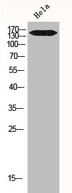
![Whole cell extract (30 μg) was separated by 5% SDS-PAGE, and the membrane was blotted with IGF1R beta antibody [HL1958] (GTX637795) diluted at 1:1000. The HRP-conjugated anti-rabbit IgG antibody (GTX213110-01) was used to detect the primary antibody.](https://www.genetex.com/upload/website/prouct_img/normal/GTX637795/GTX637795_44907_20230106_WB_R_23010922_154.webp "Whole cell extract (30 μg) was separated by 5% SDS-PAGE, and the membrane was blotted with IGF1R beta antibody [HL1958] (GTX637795) diluted at 1:1000. The HRP-conjugated anti-rabbit IgG antibody (GTX213110-01) was used to detect the primary antibody.")
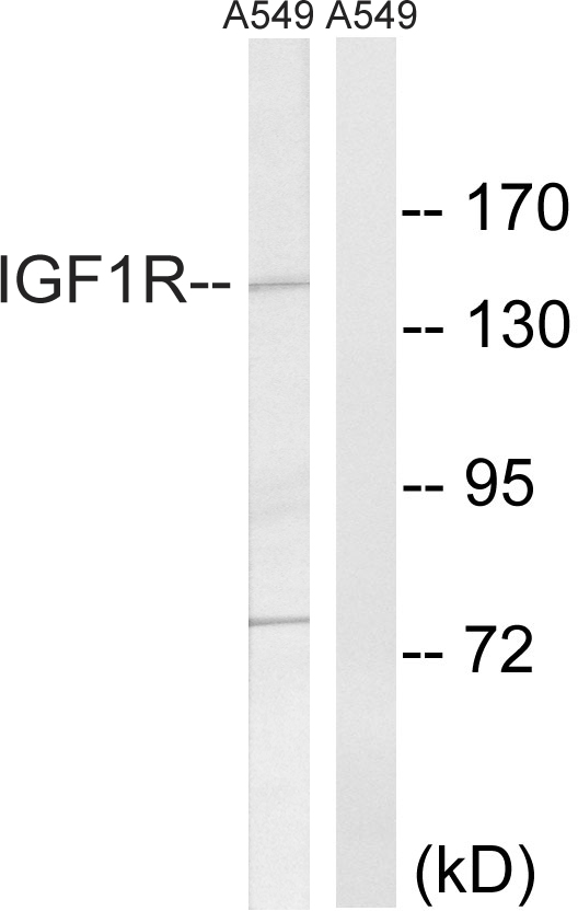
![Wild-type (WT) and IGF1R knockout (KO) HeLa cell extracts (30 μg) were separated by 5% SDS-PAGE, and the membrane was blotted with IGF1R beta antibody [HL1958] (GTX637795) diluted at 1:1000. The HRP-conjugated anti-rabbit IgG antibody (GTX213110-01) was used to detect the primary antibody.](https://www.genetex.com/upload/website/prouct_img/normal/GTX637795/GTX637795_44907_20230602_WB_KO_watermark_23060622_716.webp "Wild-type (WT) and IGF1R knockout (KO) HeLa cell extracts (30 μg) were separated by 5% SDS-PAGE, and the membrane was blotted with IGF1R beta antibody [HL1958] (GTX637795) diluted at 1:1000. The HRP-conjugated anti-rabbit IgG antibody (GTX213110-01) was used to detect the primary antibody.")
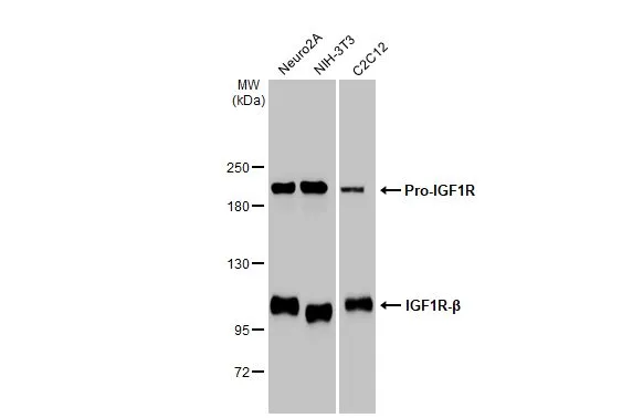
![Various whole cell extracts (30 μg) were separated by 5% SDS-PAGE, and the membrane was blotted with IGF1R beta antibody [HL1958] (GTX637795) diluted at 1:1000. The HRP-conjugated anti-rabbit IgG antibody (GTX213110-01) was used to detect the primary antibody, and the signal was developed with Trident ECL plus-Enhanced. Corresponding RNA expression data for the same cell lines are based on Human Protein Atlas program.](https://www.genetex.com/upload/website/prouct_img/normal/GTX637795/GTX637795_T-44851_20250214_WB_TPM_watermark_25021923_776.webp "Various whole cell extracts (30 μg) were separated by 5% SDS-PAGE, and the membrane was blotted with IGF1R beta antibody [HL1958] (GTX637795) diluted at 1:1000. The HRP-conjugated anti-rabbit IgG antibody (GTX213110-01) was used to detect the primary antibody, and the signal was developed with Trident ECL plus-Enhanced. Corresponding RNA expression data for the same cell lines are based on Human Protein Atlas program.")
Various whole cell extracts (30 μg) were separated by 5% SDS-PAGE, and the membrane was blotted with IGF1R beta antibody [HL1958] (GTX637795) diluted at 1:1000. The HRP-conjugated anti-rabbit IgG antibody (GTX213110-01) was used to detect the primary antibody, and the signal was developed with Trident ECL plus-Enhanced.
IGF1R beta antibody [HL1958]
GTX637795
ApplicationsWestern Blot, ImmunoHistoChemistry, ImmunoHistoChemistry Paraffin
Product group Antibodies
ReactivityHuman, Mouse, Rat
TargetIGF1R
Overview
- SupplierGeneTex
- Product NameIGF1R beta antibody [HL1958]
- Delivery Days Customer9
- Application Supplier NoteWB: 1:500-1:3000. *Optimal dilutions/concentrations should be determined by the researcher.Not tested in other applications.
- ApplicationsWestern Blot, ImmunoHistoChemistry, ImmunoHistoChemistry Paraffin
- CertificationResearch Use Only
- ClonalityMonoclonal
- Clone IDHL1958
- Concentration1 mg/ml
- ConjugateUnconjugated
- Gene ID3480
- Target nameIGF1R
- Target descriptioninsulin like growth factor 1 receptor
- Target synonymsCD221, IGFIR, IGFR, JTK13, insulin-like growth factor 1 receptor, IGF-I receptor
- HostRabbit
- IsotypeIgG
- Protein IDP08069
- Protein NameInsulin-like growth factor 1 receptor
- Scientific DescriptionThis receptor binds insulin-like growth factor with a high affinity. It has tyrosine kinase activity. The insulin-like growth factor I receptor plays a critical role in transformation events. Cleavage of the precursor generates alpha and beta subunits. It is highly overexpressed in most malignant tissues where it functions as an anti-apoptotic agent by enhancing cell survival. Alternatively spliced transcript variants encoding distinct isoforms have been found for this gene. [provided by RefSeq, May 2014]
- ReactivityHuman, Mouse, Rat
- Storage Instruction-20°C or -80°C,2°C to 8°C
- UNSPSC41116161
Datasheet
Related products
Product group Antibodies
IGF1R/INSR AntibodyCSB-PA003003
ApplicationsWestern Blot, ELISA, ImmunoHistoChemistry
ReactivityHuman, Mouse, Rat
TargetIGF1R
- SizePrice
Product group Antibodies
Igf1R Polyclonal AntibodyCAC10405
ApplicationsImmunoFluorescence, ELISA
TargetIGF1R
- SizePrice

Product group Antibodies
Anti-IGF1R Antibody Picoband(r)A00070-3-CARRIER-FREE
ApplicationsWestern Blot, ELISA
ReactivityHuman
TargetIGF1R
- SizePrice
Product group Antibodies
Anti-IGF1R AntibodyA95858
ApplicationsWestern Blot, ELISA, ImmunoHistoChemistry
ReactivityHuman, Mouse, Rat
- SizePrice
Product group Antibodies

Anti-IGF-1 [7973 [M23]], Mouse IgG1, kappaAB04263-1.1
ApplicationsWestern Blot, ImmunoHistoChemistry
ReactivityHuman, Mouse, Rat
TargetIGF1R
- SizePrice
Product group Antibodies
Anti-IGF1R Antibody144-62454
ApplicationsImmunoFluorescence, Western Blot, ImmunoHistoChemistry
ReactivityHuman, Mouse, Rat
TargetIGF1R
- SizePrice
Product group Antibodies

ApplicationsWestern Blot
ReactivityBovine, Chicken, Human, Mouse, Rat, Zebra Fish
TargetIGF1R
- SizePrice
![IGF1R beta antibody [HL1957] detects IGF1R beta protein at cell membrane by immunofluorescent analysis. Sample: MCF-7 cells were fixed in 4% paraformaldehyde at RT for 15 min. Green: IGF1R beta stained by IGF1R beta antibody [HL1957] (GTX637794) diluted at 1:1000. Blue: Fluoroshield with DAPI (GTX30920). Scale bar= 10μm.](https://www.genetex.com/upload/website/prouct_img/normal/GTX637794/GTX637794_T-44851_20230414_ICC_IF_23041719_454.webp)
Product group Antibodies
IGF1R beta antibody [HL1957]GTX637794
ApplicationsImmunoFluorescence, Western Blot, ImmunoCytoChemistry, ImmunoHistoChemistry, ImmunoHistoChemistry Paraffin
ReactivityHuman, Mouse, Rat
TargetIGF1R
- SizePrice
![IGF1R (phospho Tyr1131) / Insulin Receptor (phospho Tyr1146) antibody [HL3537] detects IGF1R (phospho Tyr1131) / Insulin Receptor (phospho Tyr1146) protein by immunofluorescent analysis. Sample: Mock and treated 293T cells were fixed in 4% paraformaldehyde at RT for 15 min. Green: IGF1R (phospho Tyr1131) / Insulin Receptor (phospho Tyr1146) stained by IGF1R (phospho Tyr1131) / Insulin Receptor (phospho Tyr1146) antibody [HL3537] (GTX641434) diluted at 1:500. Blue: Fluoroshield with DAPI (GTX30920).](https://www.genetex.com/upload/website/prouct_img/normal/GTX641434/GTX641434_T-45614_20250103_ICC_IF_treatment_IGF-1_25012200_177.webp)
Product group Antibodies
ApplicationsImmunoFluorescence, Western Blot, ImmunoCytoChemistry
ReactivityHuman
TargetIGF1R
- SizePrice